

doi: 10.1073/pnas.0405664101.
Epub 2004 Sep 7.
The inositol polyphosphate 5-phosphatase Ocrl associates with endosomes that are partially coated with clathrin
Affiliations
- PMID: 15353600
- PMCID: PMC518786
- DOI: 10.1073/pnas.0405664101
Item in Clipboard
The inositol polyphosphate 5-phosphatase Ocrl associates with endosomes that are partially coated with clathrin
Proc Natl Acad Sci U S A.
.
Abstract
The subcellular localization of Ocrl, the inositol polyphosphate 5-phosphatase that is mutated in Lowe syndrome, was investigated by fluorescence microscopy. Ocrl was localized to endosomes and Golgi membranes along with clathrin, giantin, the mannose 6-phosphate receptor, transferrin, and the early endosomal antigen 1 endosomal marker in fixed cells. The endosomal localization of Ocrl was confirmed by live-cell time-lapse microscopy in which we monitored the dynamics of Ocrl on endosomes. GST binding assays show that Ocrl interacts with the clathrin terminal domain and the clathrin adaptor protein AP-2. Our findings suggest a role for Ocrl in endosomal receptor trafficking and sorting.
Figures
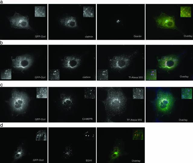
IF microscopy of Cos7 cells transiently transfected with GFP-Ocrl. Cells in a–c were serum-starved overnight in DMEM/0.1% BSA and serum-stimulated for 5 min before fixation. Cells in b and c were loaded for 30 min with 50 μg/ml Tf-Alexa 555 before serum stimulation. (a) GFP-Ocrl with clathrin and giantin stain. Overlay includes GFP-Ocrl (green), clathrin (red), and giantin (blue). (b) GFP-Ocrl-expressing cells were loaded with Tf-Alexa 555 and stained for clathrin. Overlay includes GFP-Ocrl (green), clathrin (red), and Tf (blue). (c) GFP-Ocrl-expressing cells were loaded with Tf-Alexa 555 and stained for the CI-M6PR. Overlay includes GFP-Ocrl (green), CI-M6PR (red), and Tf (blue). (d) GFP-Ocrl-expressing cells were stained for EEA1. Overlay includes GFP-Ocrl (green) and EEA1 (red).
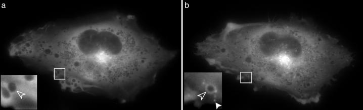
GFP-Ocrl before (a) and 23 min after (b) serum stimulation. A large increase in the number of peripheral GFP-Ocrl punctae (solid arrowhead) and rings (open arrowhead) was observed after serum stimulation. Not all vesicular structures contain Ocrl, and some acquire Ocrl upon serum stimulation (open arrowheads) (see Movie 1).
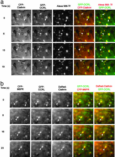
Proteins associated with Ocrl. (a) Live fluorescence microscopy of CFP-clathrin, GFP-Ocrl, and Tf-Alexa 568 after serum stimulation in Cos7 cells. The three proteins colocalize on the large vesicular structure marked by the white arrow. The open arrowhead points at a black vesicular structure that buds off a larger elongated structure. CFP-clathrin, GFP-Ocrl, and Tf-Alexa 568 are found around this budding vesicle (white arrowhead). The three proteins subsequently move with the vesicle toward the top of the picture (see Movie 3). A stationary vesicular structure is marked by an arrow. (b) Live fluorescence microscopy of CFP-CD-M6PR, GFP-Ocrl, and DsRed-clathrin after serum stimulation. Two structures that contain all three proteins are pointed out at time 0 s. The structure marked by the arrowhead then moves over the next 23 s; the other vesicular structure remains relatively stationary (see Movie 4).
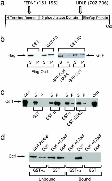
Ocrl associations. (a) Domain structure of Ocrl. Locations of the FEDNF AP-2-binding motif and the clathrin box motif LIDLE are shown. (b) Interaction between Ocrl and TD. Flag-Ocrl, GFP-Ocrl, and GFP-Ocrl LIAAA were transiently transfected into HeLa cells. GST or GST-TD beads were incubated with HeLa cell lysates. Sixteen percent of bound (P) and 2% of unbound (S) proteins were analyzed by anti-Flag Western blotting. Fourteen percent of bound (P) and 2% of unbound (S) proteins were analyzed by anti-GFP Western blotting. (c) Interaction of purified Ocrl and AP-2α. Ten percent of bound (P) and 1% of unbound (S) fractions of binding assays with GST, GST-α (AP-2α), GST-γ (AP-1γ), and GST-GGA2 (170–613) were analyzed by Ocrl Western blotting. (d) Requirement of FxDxF motif in Ocrl for AP-2 binding. Purified Ocrl or Ocrl AEANF mutant protein produced in SF9 cells was incubated with GST or GST-α appendage subunit of AP-2. Ten percent of bound and 1% of unbound fractions were analyzed by Ocrl Western blotting.
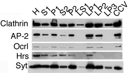
Ocrl copurifies with CCVs. Fractions were prepared as described by Maycox et al. (13). H, homogenate; S1, postnuclear supernatant; P1, nuclei/intact cells/large cell fragments; S2, supernatant minus synaptosomes; P2, crude synaptosomes; LS1, mostly cytosol; LP1, plasma membrane/coated pits/endosomes; LP2, endosomes/synaptic vesicles; LP3, CCV/remaining synaptic vesicles; CCV, highly purified CCV. The amounts of total protein loaded onto gel were as follows: H, 22.5 μg; S1, 8.25 μg; P1, 40.5 μg; S2, 6.4 μg; P2, 9 μg; LS1, 0.75 μg; LP1, 29.25 μg; LP2, 1.95 μg; LP3, 0.75 μg; CCV, 1.8 μg. The blot was probed for clathrin and Ocrl and then stripped and probed for AP-2, the multivesicular endosome marker hepatocyte growth factor-regulated tyrosine kinase substrate (Hrs), and the synaptic vesicle protein synaptotagmin (Syt).
Similar articles
-
Rab35 GTPase Triggers Switch-like Recruitment of the Lowe Syndrome Lipid Phosphatase OCRL on Newborn Endosomes.Curr Biol. 2016 Jan 11;26(1):120-8. doi: 10.1016/j.cub.2015.11.040. Epub 2015 Dec 24. Curr Biol. 2016. PMID: 26725203
-
Lowe syndrome protein OCRL1 interacts with clathrin and regulates protein trafficking between endosomes and the trans-Golgi network.Mol Biol Cell. 2005 Aug;16(8):3467-79. doi: 10.1091/mbc.e05-02-0120. Epub 2005 May 25. Mol Biol Cell. 2005. PMID: 15917292 Free PMC article.
-
Clathrin terminal domain-ligand interactions regulate sorting of mannose 6-phosphate receptors mediated by AP-1 and GGA adaptors.J Biol Chem. 2014 Feb 21;289(8):4906-18. doi: 10.1074/jbc.M113.535211. Epub 2014 Jan 9. J Biol Chem. 2014. PMID: 24407285 Free PMC article.
-
Structure and function of the Lowe syndrome protein OCRL1.Traffic. 2005 Sep;6(9):711-9. doi: 10.1111/j.1600-0854.2005.00311.x. Traffic. 2005. PMID: 16101675 Review.
-
The role of the Lowe syndrome protein OCRL in the endocytic pathway.Biol Chem. 2015 Dec;396(12):1293-300. doi: 10.1515/hsz-2015-0180. Biol Chem. 2015. PMID: 26351914 Review.
Cited by
-
Phosphoinositides: tiny lipids with giant impact on cell regulation.Physiol Rev. 2013 Jul;93(3):1019-137. doi: 10.1152/physrev.00028.2012. Physiol Rev. 2013. PMID: 23899561 Free PMC article. Review.
-
A role of the Lowe syndrome protein OCRL in early steps of the endocytic pathway.Dev Cell. 2007 Sep;13(3):377-90. doi: 10.1016/j.devcel.2007.08.004. Dev Cell. 2007. PMID: 17765681 Free PMC article.
-
The PH domain proteins IPIP27A and B link OCRL1 to receptor recycling in the endocytic pathway.Mol Biol Cell. 2011 Mar 1;22(5):606-23. doi: 10.1091/mbc.E10-08-0730. Epub 2011 Jan 13. Mol Biol Cell. 2011. PMID: 21233288 Free PMC article.
-
Suppression of intestinal calcium entry channel TRPV6 by OCRL, a lipid phosphatase associated with Lowe syndrome and Dent disease.Am J Physiol Cell Physiol. 2012 May 15;302(10):C1479-91. doi: 10.1152/ajpcell.00277.2011. Epub 2012 Feb 29. Am J Physiol Cell Physiol. 2012. PMID: 22378746 Free PMC article.
-
Impaired neural development in a zebrafish model for Lowe syndrome.Hum Mol Genet. 2012 Apr 15;21(8):1744-59. doi: 10.1093/hmg/ddr608. Epub 2011 Dec 30. Hum Mol Genet. 2012. PMID: 22210625 Free PMC article.
References
-
- Attree, O., Olivos, I. M., Okabe, I., Bailey, L. C., Nelson, D. L., Lewis, R. A., McInnes, R. R. & Nussbaum, R. L. (1992) Nature 358, 239–242. - PubMed
-
- Nussbaum, R. (2000) in The Metabolic and Molecular Bases of Inherited Disease, eds. Scriver, C. R., Sly, W.S., Childs, B., Beaudet, A. L., Valle, D., Kinzler, K. W. & Vogelstein, B. (McGraw–Hill, New York), 8th Ed., pp. 6257–6266.
-
- Majerus, P. W., Kisseleva, M. V. & Norris, F. A. (1999) J. Biol. Chem. 274, 10669–10672. - PubMed
-
- Zhang, X., Hartz, P. A., Philip, E., Racusen, L. C. & Majerus, P. W. (1998) J. Biol. Chem. 273, 1574–1582. - PubMed
-
- Suchy, S. F., Olivos-Glander, I. M. & Nussbaum, R. L. (1995) Hum. Mol. Genet. 4, 2245–2250. - PubMed
Publication types
MeSH terms
Substances
Grants and funding
LinkOut - more resources
Full Text Sources
Research Materials
