. 2003 Dec 15;17(24):3048-61.
doi: 10.1101/gad.1153003.

SUMO modification of a novel MAR-binding protein, SATB2, modulates immunoglobulin mu gene expression

Affiliations

SUMO modification of a novel MAR-binding protein, SATB2, modulates immunoglobulin mu gene expression

Gergana Dobreva et al. Genes Dev. .

Abstract

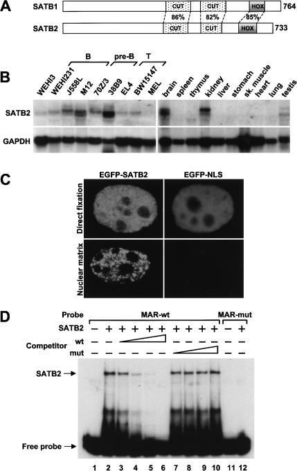
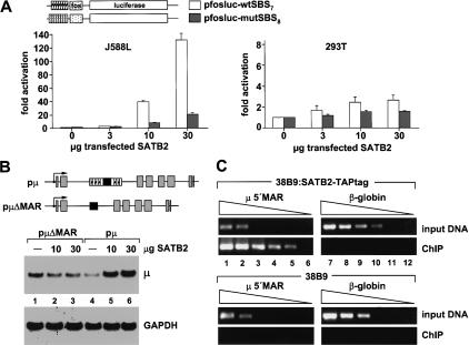
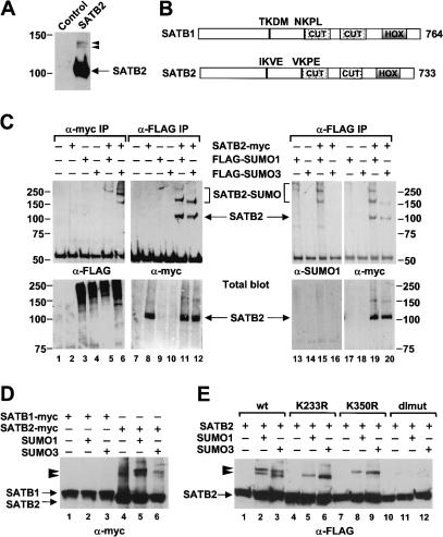
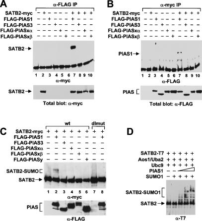
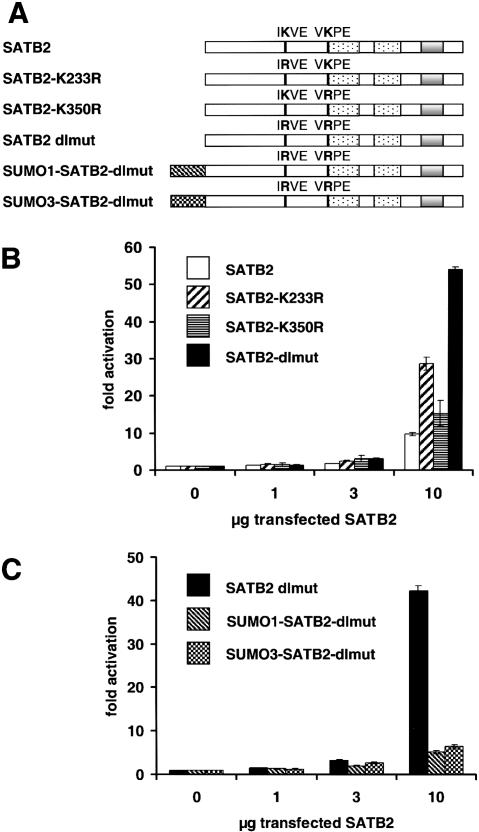
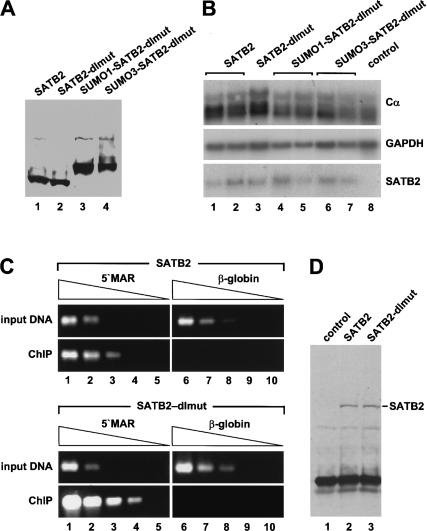
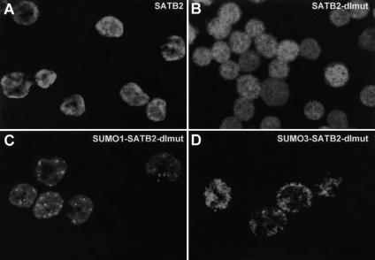
Nuclear matrix attachment regions (MARs) are regulatory DNA sequences that are important for higher-order chromatin organization, long-range enhancer function, and extension of chromatin modifications. Here we characterize a novel cell type-specific MAR-binding protein, SATB2, which binds to the MARs of the endogenous immunoglobulin micro locus in pre-B cells and enhances gene expression. We found that SATB2 differs from the closely related thymocyte-specific protein SATB1 by modifications of two lysines with the small ubiquitive related modifier (SUMO), which are augmented specifically by the SUMO E3 ligase PIAS1. Mutations of the SUMO conjugation sites of SATB2 enhance its activation potential and association with endogenous MARs in vivo, whereas N-terminal fusions with SUMO1 or SUMO3 decrease SATB2-mediated gene activation. Sumoylation is also involved in targeting SATB2 to the nuclear periphery, raising the possibility that this reversible modification of a MAR-binding protein may contribute to the modulation of subnuclear DNA localization.


Figures

Figure 1.
Figure 1.
Cell type-specific expression and nuclear matrix association of SATB2. (A) Schematic representation of the domain structure of SATB1 and SATB2. The amino acid sequence identities in the MAR-binding domain (CUT domains) and the homeodomain (HOX) are indicated in percentages. (B) RNA blot analysis of 20 μg total RNA isolated from various B- and T-lymphoid cell lines, a murine erythroid (MEL) and myeloid (WEHI3) cell line (left-hand panels), and from various mouse tissues (right-hand panels). Abundant expression of ∼6.3- and ∼5.4-kb transcripts that hybridize with a 32P-labeled satb2 DNA probe can be detected in B cells, brain, and kidney (top panels). The quality and quantity of RNA were confirmed by hybridization with a GAPDH probe (bottom panels). (C) Association of SATB2 with the nuclear matrix. 293T cells were transfected with an EGFP-SATB2 or EGFP-NLS gene construct, and after 48 h the fusion proteins were visualized by fluorescence microscopy in directly fixed cells (top panels) or in nuclear matrix preparations (bottom panels). (D) EMSA to detect binding of purified recombinant SATB2 (30 ng) to 32P-labeled wild-type (wt) or mutated (mut) MAR oligonucleotides (lanes 2,12). The specificity of DNA binding by SATB2 was shown by competition with increasing amounts of unlabeled wild-type or mutant MAR oligonucleotides (lanes 3-10).
Figure 2.
Figure 2.
SATB2 augments μ gene expression and binds to endogenous MAR sequences of the μ locus. (A) SATB2 stimulates a luciferase reporter gene containing multimerized SATB2-binding sites in J558L plasmacytoma cells but not in 293T cells. Cells were transfected with 5 μg reporter construct, together with 1 μg β-galactosidase reporter construct for normalization and increasing amounts of SATB2 expression plasmid, as indicated. The levels of luciferase activity, normalized to the activity of the cotransfected β-galactosidase reporter, are expressed as fold-activation relative to the luciferase levels of cells transfected with the reporters alone. All transfections were performed at least three times, and representative experiments with the standard deviations are shown. (B) SATB2 augments the expression of a rearranged μ immunoglobulin gene in a MAR-dependent manner. J558L cells were transfected with 5 μg of wild-type or μΔMAR genes and increasing amounts of SATB2 expression plasmid (10, 30 μg), as indicated. Expression of the μ gene was examined by RNA blot analysis of total RNA (20 μg) with a 32P-labeled VH17.2.25-specific DNA probe (top panel) or GAPDH probe (bottom panel). (C) SATB2 binds to the MAR of the endogenous μ locus. ChIP of extracts from stably transfected 38B9 cells carrying a SATB2-TAPtag expression plasmid, or from control 38B9 cells. Chromatin fragments that had been cross-linked to SATB2-TAPtag were affinity-purified, and the immunoprecipitated DNA was subsequently analyzed by semiquantitative PCR amplification with primers located in the 5′ MAR region of the immunoglobulin μ enhancer (lanes 1-6) or in the β-globin locus (lanes 7-12). The levels of enrichment in the immunoprecipitations were estimated by comparison with the amplification products of DNA isolated from the bulk chromatin extracts (input DNA). Template DNA was used in a linear dilution (3, 1, 0.3, 0.1, 0.03, 0.01 ng) to allow for a semiquantitative determination in the PCR assays.
Figure 3.
Figure 3.
SATB2 is SUMO-modified in vivo. (A) Immunoblot analysis of total protein extracts from 293T cells transfected transiently with a myc-tagged SATB2 expression plasmid or a control vector. In addition to SATB2, migrating at ∼105 kD, two forms of SATB2 migrating at ∼135 kD and ∼140 kD can be detected with an anti-myc antibody. (B) Schematic representation of the domain structure of SATB1 and SATB2. The two CUT motifs, the homeobox (HOX), and the amino acid sequence of two putative SUMO acceptor sites at positions 233 (IKVE) and 350 (VKPE) of SATB2 are shown. The corresponding sequences in SATB1 do not conform to the consensus SUMO acceptor site ψKXE. (C) Modification of SATB2 by SUMO1 and SUMO3. Myc-tagged SATB2 and Flag-tagged SUMO1 or SUMO3 expression plasmids were transiently transfected into 293T cells. Equivalent amounts (500 μg) of total cellular protein were immunoprecipitated with an anti-myc antibody (lanes 1-6, top panel) or an anti-Flag antibody (lanes 7-20, top panels), and the immunoprecipitated proteins that have been modified with Flag-SUMO proteins were detected with an anti-Flag antibody (lanes 1-6) or an anti-SUMO1 antibody (lanes 13-16). SATB2 and SUMO-modified forms of SATB2 were detected with an anti-myc antibody (lanes 7-12 and 17-20, top panels). Similar expression of SATB2-myc, Flag-SUMO1, and Flag-SUMO3 was confirmed by immunoblot analysis of total cell lysates (bottom panels). (D) SATB2, but not SATB1, can be modified with SUMO proteins. Myc-tagged SATB1 (lanes 1-3) and myc-tagged SATB2 (lanes 4-6) are detected in total cell lysates of transfected cells by an anti-myc immunoblot analysis. SUMO-modified forms of SATB2 are marked by arrowheads. (E) Identification of K233 and K350 of SATB2 as the acceptor sites for sumoylation. Total protein extracts from cells transfected with expression plasmids of Flag-SATB2 (lanes 1-3) or various Flag-tagged SATB2 mutants: K233R (lanes 4-6), K350R (lanes 7-9), or the K233R/K350R double mutant (dlmut, lanes 10-12), alone or together with SUMO1 or SUMO3 expression plasmids, were analyzed by an immunoblot with an anti-Flag antibody. Mutation of either sumoylation site interfered with the appearance of one of the two modified forms of SATB2, whereas the double mutation abrogated SUMO modification. The differences in the migration of the two modified forms of SATB2 are most likely due to the branching position of the sumoylated polypeptides (Hoege et al. 2002).
Figure 4.
Figure 4.
PIAS1 interacts with SATB2 and stimulates SUMO modification of SATB2 in vivo and in vitro. (A,B) PIAS1 interacts with SATB2 in vivo. Myc-tagged SATB2 and Flag-tagged forms of PIAS1, PIAS3, PIASxα, and PIASxβ were transiently expressed in 293T cells. Equivalent amounts of total cellular protein were immunoprecipitated with an anti-Flag antibody (A, lanes 1-10) or an anti-myc antibody (B, lanes 1-10). The co-immunoprecipitated proteins were detected by an immunoblot analysis with an anti-myc antibody (A, lanes 1-10, top panel) or anti-Flag antibody (B, lanes 1-10, top panel). Similar expression of SATB2 and the PIAS proteins was confirmed by immunoblot analysis of total cell lysates (bottom panels). (C) PIAS1 stimulates SUMO conjugation to SATB2 in vivo. Myc-tagged SATB2 or SATB2-dlmut were expressed alone or together with Flag-tagged PIAS proteins in 293T cells. SATB2 and SUMO-modified SATB2 were detected in total cell lysates by immunoblot analysis with an anti-myc antibody. SUMO-modified SATB2 can be detected only in cells expressing PIAS1 (lane 2). (D) PIAS1 stimulates SUMO conjugation to SATB2 in vitro: 300 ng purified His- and T7-double-tagged SATB2 protein was incubated for 30 min with 250 ng purified E1 enzyme (Aos1/Uba2 heterodimer), 250 ng E2 enzyme (Ubc9), and 1 μg SUMO1, alone or with increasing amounts of GST-PIAS1. SATB2 proteins were detected by an anti-T7 immunoblot analysis.
Figure 5.
Figure 5.
Sumoylation antagonizes SATB2-mediated transcriptional activation. (A) Schematic representation of wild-type SATB2 and mutant SATB2 proteins containing mutations of the sumoylation sites, individually (K233R, K350R) or in combination (dlmut), and carrying an amino-terminal fusion of SUMO1 (SUMO1-SATB2-dlmut) or SUMO3 (SUMO3-SATB2-dlmut). The amino-terminal fusions of SUMO cannot be hydrolyzed by SUMO isopeptitases. (B) Mutation of both sumoylation sites of SATB2 augments transcriptional activation. J558L cells were transiently transfected by electroporation with 5 μg of a luciferase reporter construct containing multimerized SATB2-binding sites, alone or together with increasing amounts (1, 3, and 10 μg) of expression plasmids encoding wild-type SATB2 or mutated SATB2 proteins. For the normalization of luciferase activities, the activity of a cotransfected β-galactosidase expression plasmid (1 μg) was determined for each sample. The normalized levels of luciferase activity are expressed as fold-activation relative to the level of luciferase activity from cells transfected with the reporter construct alone. (C) Amino-terminal fusions of SUMO1 or SUMO3 antagonize SATB2-mediated transcriptional activation. J558L cells were transiently transfected by electroporation with a luciferase reporter construct, alone or together with increasing amounts of expression plasmids encoding SATB2-dlmut, SUMO1-SATB2-dlmut, or SUMO3-SATB2-dlmut as indicated.
Figure 6.
Figure 6.
Mutations of the sumoylation sites of SATB2 augment the association with MAR sequences of the endogenous immunoglobulin heavy-chain locus. (A) Immunoblot analysis of clones of J558L plasmacytoma cells that have been stably transfected with Flag-tagged wild-type SATB2 and various mutant SATB2 expression plasmids under the control of the EF1α promoter. (B) RNA blot analysis of J558L clones that have been stably transfected with various SATB2 constructs. Five micrograms total RNA was hybridized with probes that detect the endogenous Cα and GAPDH transcripts and the transfected satb2 mRNA. (C) ChIP experiments of J558L cell lines expressing similar amounts of Flag-tagged wild-type SATB2 (top panels) or the sumoylation site-deficient SATB2 double mutant (bottom panels). DNA of chromatin fragments that had been immunoprecipitated with an anti-Flag antibody was subsequently analyzed by semiquantitative PCR, using primers located in the 5′ MAR region of the immunoglobulin μ enhancer (lanes 1-5) or in the β-globin locus (lanes 6-10). Template DNA was used in a fivefold linear dilution series (starting from 5 ng). (D) Anti-Flag immunoblot analysis to detect SATB2 proteins that have been immunoprecipitated under ChIP conditions to control for equal fractionation and recovery of SATB2 and SATB2-dlmut.
Figure 7.
Figure 7.
SUMO modification alters the subnuclear localization of SATB2. J558L plasmacytoma cells were stably transfected with plasmids encoding Flag-tagged wild-type and mutant SATB2 proteins, and clones expressing similar amounts of SATB2 protein were chosen for the analysis of the subcellular localization by indirect immunofluorescence (Fig. 6A). (A) Wild-type SATB2 localizes predominantly to the nuclear periphery. (B) SATB2-dlmut is diffusely distributed throughout the nucleus. (C) Covalent attachment of SUMO1 to the amino terminus of SATB2-dlmut localizes the protein into nuclear bodies. (D) Covalent attachment of SUMO3 to the amino terminus of SATB2-dlmut localizes the protein predominantly to the nuclear periphery.

Similar articles

Cited by

References

    1. Alvarez J.D., Yasui, D.H., Niida, H., Joh, T., Loh, D.Y., and Kohwi-Shigematsu, T. 2000. The MAR-binding protein SATB1 orchestrates temporal and spatial expression of multiple genes during T-cell development. Genes & Dev. 14: 521-535. - PMC - PubMed
    1. Berezney R. and Coffey, D.S. 1974. Identification of a nuclear protein matrix. Biochem. Biophys. Res. Commun. 60: 1410-1417. - PubMed
    1. Best J.L., Ganiatsas, S., Agarwal, S., Changou, A., Salomoni, P., Shirihai, O., Meluh, P.B., Pandolfi, P.P., and Zon, L.I. 2002. SUMO-1 protease-1 regulates gene transcription through PML. Mol. Cell 10: 843-855. - PubMed
    1. Bode J., Benham, C., Knopp, A., and Mielke, C. 2000. Transcriptional augmentation: Modulation of gene expression by scaffold/matrix-attached regions (S/MAR elements). Crit. Rev. Eukaryot. Gene Expr. 10: 73-90. - PubMed
    1. Cai S., Han, H.J., and Kohwi-Shigematsu, T. 2003. Tissue-specific nuclear architecture and gene expression regulated by SATB1. Nat. Genet. 34: 42-51. - PubMed

Publication types

MeSH terms

Substances

Associated data