. 2003 Jun 1;23(11):4549-59.
doi: 10.1523/JNEUROSCI.23-11-04549.2003.

Connexin 47 (Cx47)-deficient mice with enhanced green fluorescent protein reporter gene reveal predominant oligodendrocytic expression of Cx47 and display vacuolized myelin in the CNS

Affiliations

Connexin 47 (Cx47)-deficient mice with enhanced green fluorescent protein reporter gene reveal predominant oligodendrocytic expression of Cx47 and display vacuolized myelin in the CNS

Benjamin Odermatt et al. J Neurosci. .

Abstract

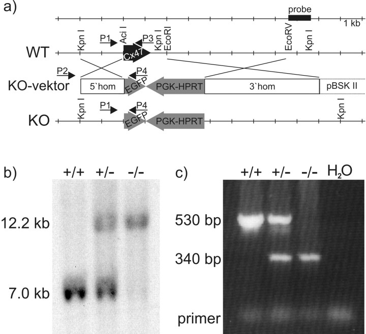
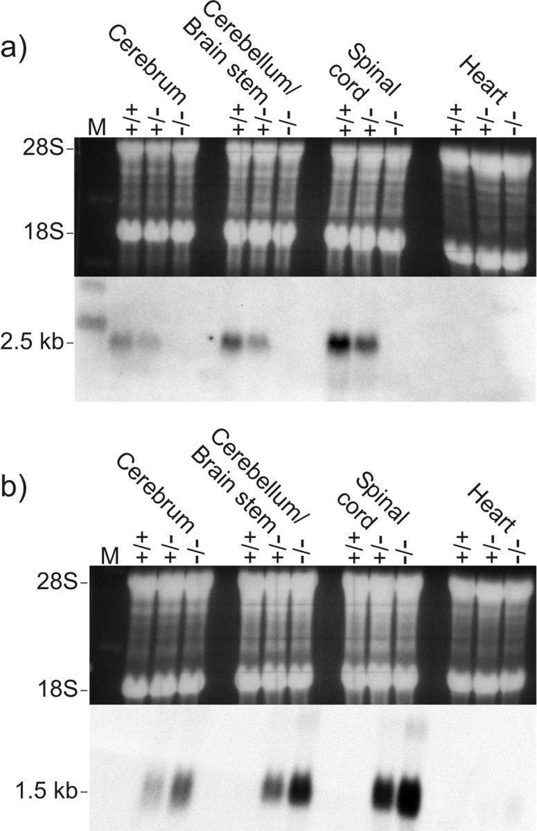
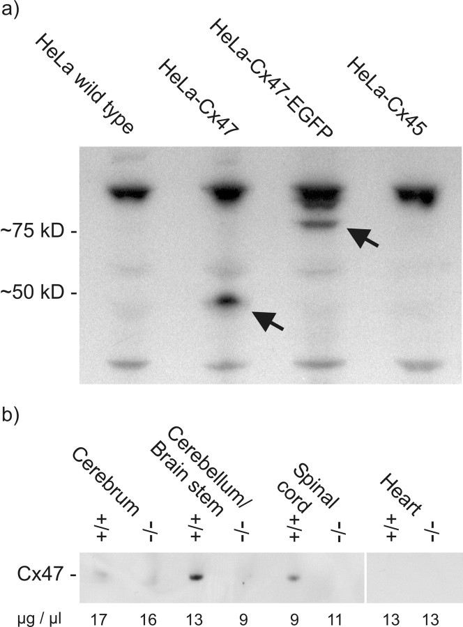
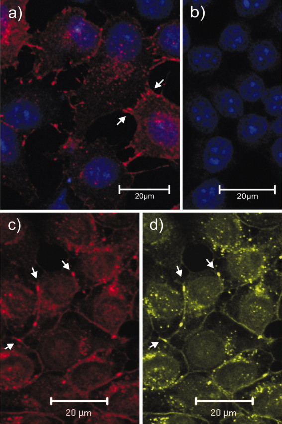
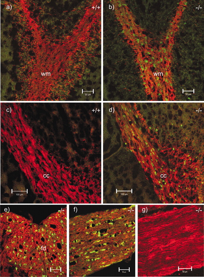
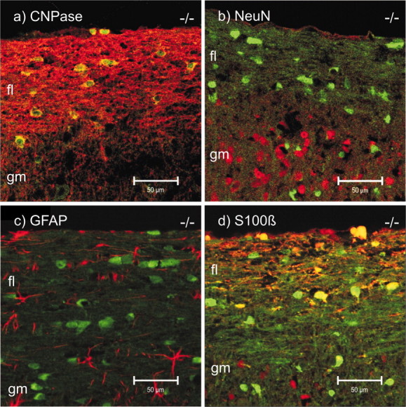
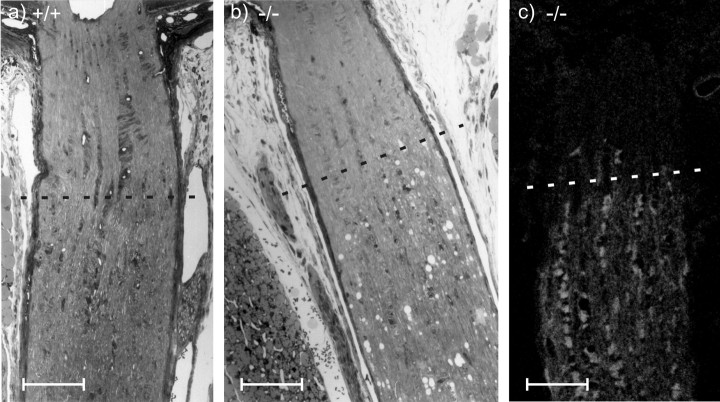
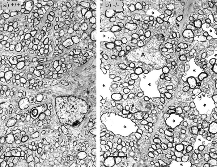
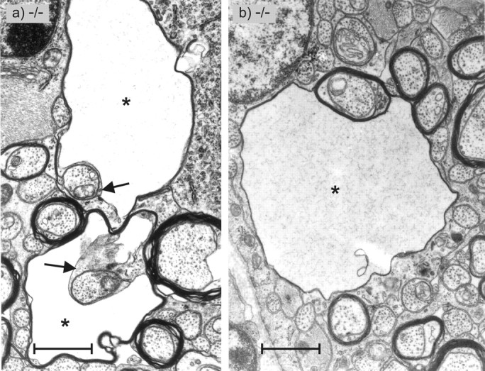
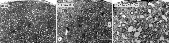
To further characterize the recently described gap junction gene connexin 47 (Cx47), we generated Cx47-null mice by replacing the Cx47 coding DNA with an enhanced green fluorescent protein (EGFP) reporter gene, which was thus placed under control of the endogenous Cx47 promoter. Homozygous mutant mice were fertile and showed no obvious morphological or behavioral abnormalities. Colocalization of EGFP fluorescence and immunofluorescence of cell marker proteins revealed that Cx47 was mainly expressed in oligodendrocytes in highly myelinated CNS tissues and in few calcium-binding protein S100beta subunit-positive cells but not in neurons or peripheral sciatic nerve. This corrects our previous conclusion that Cx47 mRNA is expressed in brain and spinal cord neurons (Teubner et al., 2001). Cx47 protein was detected by Western blot analysis after immunoprecipitation in CNS tissues of wild-type mice but not in heart or Cx47-deficient tissues. Electron microscopic analysis of CNS white matter in Cx47-deficient mice revealed a conspicuous vacuolation of nerve fibers, particularly at the site of the optic nerve where axons are first contacted by oligodendrocytes and myelination starts. Initial analyses of Cx32/Cx47-double-deficient mice showed that these mice developed an action tremor and died on average at 51 d after birth. The central white matter of these double-deficient mice exhibited much more abundant vacuolation in nerve fibers than mice deficient only in Cx47.


Figures

Figure 1.
Figure 1.
Generation of Cx47-deficient mice with the EGFP reporter gene (Cx47 EGFP). a, Targeting scheme. WT, Wild-type allele; KO, knock-out allele. The coding region of the wild-type Cx47 gene (black arrow) is located on one exon and includes 1314 bp. After electrotrans-fecting HM-1 cells with the linearized targeting vector (KO-vektor), homologous recombination occurred in 10% of all clones as determined by PCR using the external sense primer (P2) and the EGFP-specific antisense primer (P4). In the mutated Cx47 allele, the coding region of Cx47 after the first seven codons was replaced by the EGFP cDNA and the PGK-HPRT minigene (gray arrows). The translational start codon of EGFP was cloned in-frame with the Cx47 coding region. The PGK-HPRT minigene was transcribed in the opposite direction. The backbone of the targeting vector consisted of pBlueskript SK II(+) (pBSK II; Stratagene). The 5′ homologous region (5′hom) spanned 2 kb; the 3′ homologous region (3′hom) included 5.5 kb. b, Southern blot analysis was performed to confirm homologous recombination. KpnI-digested mouse kidney DNA was probed with a 1 kb external EcoRV–KpnI fragment (probe). The 7.0 kb band was derived from the wild-type allele; the larger 12.2 kb band resulted from the knock-out allele. c, Diagnostic PCR of tail-tip DNA was performed to screen pedigree mice for occurrence of wild-type and knock-out Cx47 EGFP alleles. A multiplex PCR was established using a Cx47 intron-specific sense primer (P1) and a Cx47 exon-specific antisense primer (P3), producing a 530 bp wild-type amplicon and an EGFP-specific antisense primer (P4), resulting in a 340 bp amplicon of the knock-out allele.
Figure 2.
Figure 2.
Northern blot analysis of Cx47 expression (a) and EGFP expression (b) in adult mouse tissues. Equal amounts of total RNA (20 μg) from wild-type (+/+), heterozygous (+/-), and homozygous (-/-) Cx47 EGFP mice (littermates) were applied as demonstrated by staining of 28S and 18S ribosomal RNA with ethidium bromide (top). The blotted RNA (bottom) was probed with a 1.6 kb XbaI fragment containing part of the Cx47 coding region and 3′-untranslated region (a) and a 1.0 kb SalI–AflII fragment of the pEGFP-1 vector containing the EGFP-coding region and the SV40 poly(A) signal (b). The size of the corresponding transcripts is indicated in kilobases on the left.
Figure 3.
Figure 3.
Western blot analysis of protein lysates from different HeLa connexin transfectants (a) and after immunoprecipitation of protein lysates from Cx47 wild-type (+/+) and Cx47 EGFP(-/-) mouse tissues (b). Western blot analysis with Cx47 antibodies detected by 125I-labeled protein A showed specific bands, at ∼50 kDa in HeLa Cx47 lysates and ∼75 kDa in HeLaCx47-EGFPlysates. Nonspecific bands at ∼30 and 100 kDa also appeared in control lysates of HeLa wild-type cells and Cx45 transfectants (a). Molecular masses are indicated in kilodaltons on the left. By Western blot analysis (ECL) with biotinylated Cx47 antibodies after immunoprecipitation with Cx47 antibodies in lysates of mouse tissue (b), Cx47 was found in the cerebellum and spinal cord of wild-type mice; in cerebrum, the signal was very weak. In heart (control tissue) and lysates from Cx47 EGFP(-/-) littermates, no bands were detected. Numbers under each lane represent micrograms of total protein per microliter of lysate of the tissue indicated.
Figure 4.
Figure 4.
Immunostaining (red) of Cx47 in HeLa-Cx47 (a), HeLa wild-type (b), and HeLa-Cx47-EGFP (c) cells. The EGFP fluorescent signals (d, green) corresponding to Cx47-EGFP fusion proteins were primarily colocalized with Cx47 immunofluorescent signals (c). Typical punctate gap junction staining was detected on contact membranes of Cx47-transfected cells (arrows), whereas no such staining could be detected in HeLa wild-type controls. Nuclei in HeLa-Cx47 and HeLa wild-type cells were stained with TOTO-1 (blue, false color); nuclei in HeLa-Cx47-EGFP cells were not stained but are visible because of higher background staining. Scale bars, 20 μm.
Figure 5.
Figure 5.
EGFP signal (green) as reporter for Cx47-expressing cells and immunostaining (red) for MBP in wild-type (+/+) and homozygous Cx47 EGFP (-/-) mice. Shown are double (red, green) confocal images of cerebellar white matter (wm; a, b), corpus callosum (cc; c, d), the funiculus dorsalis (fd) in a transverse section of the spinal cord (e), a longitudinal section of the optic nerve (f), and a longitudinal section of the sciatic nerve (g). All corresponding micrographs (a–g) were recorded with the same microscope settings. No EGFP signal was observed in wild-type tissues (a, c) and Cx47 EGFP(-/-) sciatic nerve (g). Scale bars: a, b, e–g, 50 μm; c, d, 100 μm.
Figure 6.
Figure 6.
EGFP signal (green) as reporter for Cx47-expressing cells and immunostaining (red) for oligodendrocytic CNPase (a), NeuN (b), GFAP (c), and S100β in longitudinal sections of the spinal cord of Cx47 EGFP(-/-) mice. Double (red, green) confocal images are shown. All corresponding images (a–d) were recorded with the same microscope settings. CNPase colocalized (yellow) with EGFP fluorescence in the pericaryon (a). Cells immunolabeled for NeuN (b) were EGFP-negative. Likewise, GFAP-positive cells (c) did not express EGFP. A few S100β-positive cells (d) expressed EGFP (yellow); others did not (red), but most EGFP-expressing cells were negative for S100β (green). Scale bars, 50 μm. fl, Funiculus lateralis; gm, gray matter.
Figure 7.
Figure 7.
Electrophysiological properties of GFP-labeled cells in Cx47 EGFP(+/-) and PLP-GFP mice. Membrane currents were activated between -160 and +20 mV (50 msec; 10 mV increment; holding potential, -70 mV). a, b, Right, I–V plots when taking amplitudes 2 msec (triangles) and 50 msec (diamonds) after the onset of the voltage steps. a, In a fluorescent cell, presumed an oligodendrocyte of the corpus callosum of a Cx47 EGFP(+/-) mouse (resting potential, -60 mV), currents reversed at -67 and -59 mV, respectively. b, A GFP-positive oligodendrocyte in the corpus callosum of a PLP-GFP mouse (resting potential, -70 mV) displayed reversal potentials of -67 and -64 mV. In both cells, currents decayed with similar time constants as indicated (V, +10 mV; a single exponential was fit to the data recorded between 3 and 25 msec after onset of the voltage steps ; time window marked by dashed vertical bars). c, Subsequent to functional characterization, the cytoplasm of the cells shown in a and b was harvested for transcript analysis. PCR products specific to Cx47 (334 bp) were identified by gel electrophoresis (lanes #1, #2 correspond to the cells in a, b, respectively). Cx47 cDNA was completely digested by the restriction endonuclease MboI, yielding fragments of 239 and 95 bp (lanes #1c, #2c). d, A GFP-negative CA1 pyramidal neuron (lane #3; PLP-GFP mouse) and an EGFP-negative astrocyte (lane #4; CA1 stratum oriens, Cx47 EGFP(+/-) mouse) were analyzed as described above. Both cells lacked Cx47 transcripts, although mRNA of the housekeeping gene β-actin (238 bp) was found (right). As a length marker, HincII-digested DNA from φX174 phages was used.
Figure 8.
Figure 8.
Longitudinal semithin sections of the optic nerve, close to the eye. The partly myelinated transition zone of the optic nerve starts ∼250 μm behind the eye bulb (dashed line). In contrast to the wild-type tissue (a), the myelinated region of the optic nerve from Cx47 EGFP(-/-) mice showed many vacuolated nerve fibers (b). Confocal microscopic analysis of EGFP fluorescence (light gray) in the same region of Cx47 EGFP(-/-) mice (c) demonstrated that Cx47 expression did not occur in front of this transition zone. Scale bars, 100 μm.
Figure 9.
Figure 9.
Transverse sections through the partly myelinated transition zone of the optic nerve. In wild-type tissue (a) as well as in Cx47 EGFP(-/-) tissue (b), thin myelin sheets of the first internodes and nonmyelinated nerve fibers were seen. In this transition zone, the vacuolated nerve fibers of the Cx47 EGFP(-/-) animal were very prominent (asterisks). A, Astrocytes. Scale bars, 3.6 μm.
Figure 10.
Figure 10.
Transverse sections through the partly myelinated transition zone of the optic nerve of Cx47 EGFP(-/-) mice as shown in Figure 9. Two types of vacuolated nerve fibers are shown. a, The two nerve fibers exhibit vacuoles (asterisks) between the inner lamellae of the oligodendrocyte (arrows) and the compact lamellae of the myelin sheet. b, The vacuole (asterisk) of this nerve fiber is located between compact myelin sheets. Scale bars, 1.2 μm.
Figure 11.
Figure 11.
Transverse semithin sections through the intracranial part of the optic nerve immediately in front of the optic chiasm of 4-week-old Cx32 (y/+)/Cx47 (+/+) wild-type (wt; a), 5-week-old Cx47 EGFP(-/-) single-deficient (b), and a Cx32 (y/-)/Cx47 EGFP(-/-) double-deficient (c) mice (b, c are littermates). The double-deficient mice exhibited strongly enhanced vacuolation of nerve fibers all over the CNS, whereas the Cx47 EGFP(-/-) mice showed very rare vacuolation (asterisks). Cx47 EGFP(-/-) mice exhibited increased vacuolation only in the transition zone of the optic nerve (Fig. 9b). KO, Knock-out. Scale bars, 16 μm.

Similar articles

Cited by

References

    1. Altevogt BM, Kleopa KA, Postma FR, Scherer SS, Paul DL ( 2002) Connexin29 is uniquely distributed within myelinating glial cells of the central and peripheral nervous systems. J Neurosci 22: 6458–6470. - PMC - PubMed
    1. Anzini P, Neuberg DH, Schachner M, Nelles E, Willecke K, Zielasek J, Toyka KV, Suter U, Martini R ( 1997) Structural abnormalities and deficient maintenance of peripheral nerve myelin in mice lacking the gap junction protein connexin 32. J Neurosci 17: 4545–4551. - PMC - PubMed
    1. Barger SW, Wolchok SR, Van Eldik LJ ( 1992) Disulfide-linked S100 beta dimers and signal transduction. Biochim Biophys Acta 1160: 105–112. - PubMed
    1. Berger T, Schnitzer J, Kettenmann H ( 1991) Developmental changes in the membrane current pattern, K+ buffer capacity, and morphology of glial cells in the corpus callosum slice. J Neurosci 11: 3008–3024. - PMC - PubMed
    1. Bosio A, Büssow H, Adam J, Stoffel W ( 1998) Galactosphingolipids and axono-glial interaction in myelin of the central nervous system. Cell Tissue Res 292: 199–210. - PubMed

Publication types

MeSH terms