

New syndrome characterized by hypomyelination with atrophy of the basal ganglia and cerebellum
- PMID: 12372733
- PMCID: PMC7976795
New syndrome characterized by hypomyelination with atrophy of the basal ganglia and cerebellum
Abstract
Background and purpose: Leukoencephalopathies of unknown origin constitute a considerable problem in child neurology. The purpose of our ongoing study of the subject was to define new disease entities among them by using primarily MR imaging pattern recognition.
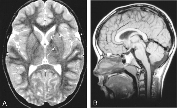
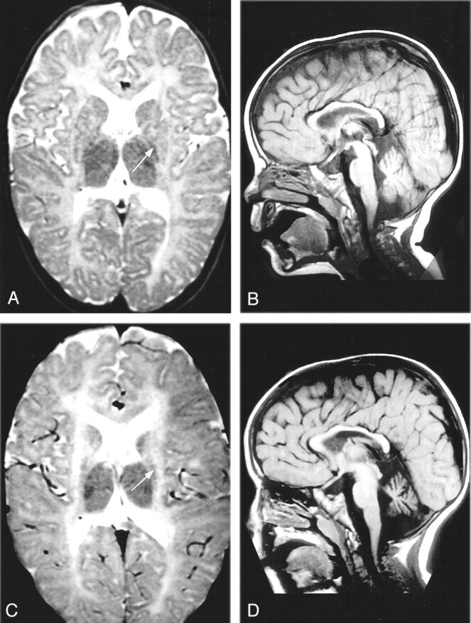
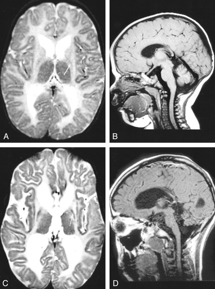
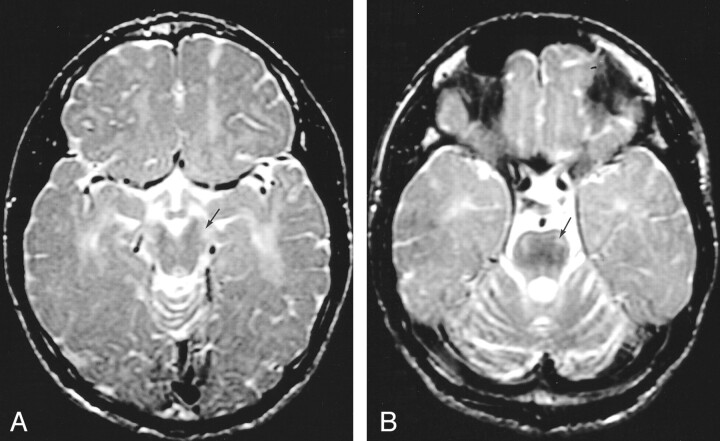
Methods: We identified seven unrelated patients with a distinct MR imaging pattern consisting of hypomyelination and atrophy of the basal ganglia (neostriatum) and cerebellum (H-ABC). We reviewed the clinical, MR imaging, MR spectroscopic, and laboratory data.
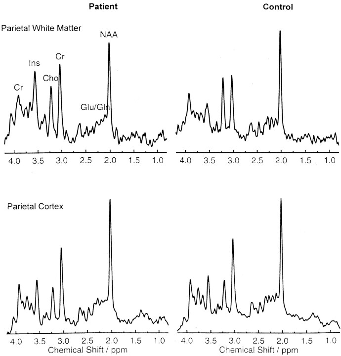
Results: Clinically, the patients' diseases were characterized by variably disturbed early development followed by increasing extrapyramidal movement abnormalities, ataxia, and spasticity. Mental capacity was variably affected, but it appeared to be relatively preserved. Parents were not related, and none of their siblings were affected. No metabolic defect was found. Follow-up MR imaging demonstrated atrophy of the cerebral white matter, neostriatum, and cerebellum, which was most pronounced in the most clinically severe cases. Single-voxel proton MR spectroscopic results were normal in the parietal cortex. In the cerebral white matter, myo-inositol and creatine levels were elevated; this finding was compatible with gliosis. N-acetylaspartate and choline levels were normal, suggesting that neither axonal loss nor active demyelination occurred. Proton MR spectroscopic imaging revealed relatively decreased N-acetylaspartate levels in the frontal region.
Conclusion: The uniform and highly characteristic MR imaging findings, in combination with the similarities in the clinical findings, provide evidence of a distinct nosologic entity. The acronym H-ABC is offered to indicate patients sharing these clinical and MR imaging features.
Figures
Similar articles
-
Hypomyelination with atrophy of the basal ganglia and cerebellum: further delineation of the phenotype and genotype-phenotype correlation.Brain. 2014 Jul;137(Pt 7):1921-30. doi: 10.1093/brain/awu110. Epub 2014 Apr 30. Brain. 2014. PMID: 24785942 Free PMC article.
-
Hypomyelination with atrophy of the basal ganglia and cerebellum: follow-up and pathology.Neurology. 2007 Jul 10;69(2):166-71. doi: 10.1212/01.wnl.0000265592.74483.a6. Neurology. 2007. PMID: 17620549
-
[A boy with hypomyelination with atrophy of the basal ganglia and cerebellum].No To Hattatsu. 2010 Jan;42(1):42-4. No To Hattatsu. 2010. PMID: 23858611 Japanese.
-
Novel TUBB4A mutations and expansion of the neuroimaging phenotype of hypomyelination with atrophy of the basal ganglia and cerebellum (H-ABC).Am J Med Genet A. 2014 Jul;164A(7):1802-7. doi: 10.1002/ajmg.a.36526. Epub 2014 Apr 4. Am J Med Genet A. 2014. PMID: 24706558 Free PMC article. Review.
-
[A report of atypical hypomyelinating leukodystrophy with atrophy of the basal ganglia and cerebellum caused by a de novo mutation in tubulin beta 4A (TUBB4A) gene and literature review].Zhonghua Nei Ke Za Zhi. 2017 Jun 1;56(6):433-437. doi: 10.3760/cma.j.issn.0578-1426.2017.06.009. Zhonghua Nei Ke Za Zhi. 2017. PMID: 28592043 Review. Chinese.
Cited by
-
Hypomyelinating leukoencephalopathy.Sultan Qaboos Univ Med J. 2013 Feb;13(1):192-3. Epub 2013 Feb 27. Sultan Qaboos Univ Med J. 2013. PMID: 23573409 Free PMC article. No abstract available.
-
Hypomyelination and congenital cataract: neuroimaging features of a novel inherited white matter disorder.AJNR Am J Neuroradiol. 2008 Feb;29(2):301-5. doi: 10.3174/ajnr.A0792. Epub 2007 Nov 1. AJNR Am J Neuroradiol. 2008. PMID: 17974614 Free PMC article.
-
Emerging cellular themes in leukodystrophies.Front Cell Dev Biol. 2022 Aug 8;10:902261. doi: 10.3389/fcell.2022.902261. eCollection 2022. Front Cell Dev Biol. 2022. PMID: 36003149 Free PMC article. Review.
-
Mutations in POLR3A and POLR3B encoding RNA Polymerase III subunits cause an autosomal-recessive hypomyelinating leukoencephalopathy.Am J Hum Genet. 2011 Nov 11;89(5):644-51. doi: 10.1016/j.ajhg.2011.10.003. Epub 2011 Oct 27. Am J Hum Genet. 2011. PMID: 22036171 Free PMC article.
-
Neurological Disorders Associated with Striatal Lesions: Classification and Diagnostic Approach.Curr Neurol Neurosci Rep. 2016 Jun;16(6):54. doi: 10.1007/s11910-016-0656-3. Curr Neurol Neurosci Rep. 2016. PMID: 27074771 Review.
References
-
- Van der Knaap MS, Breiter SN, Naidu S, et al. Defining and categorizing leukoencephalopathies of unknown origin: MR imaging approach. Radiology 1999;213:121–133 - PubMed
-
- Kristjánsdóttir R, Uvebrant P, Hagberg B, et al. Disorders of the cerebral white matter in children: The spectrum of lesions. Neuropediatrics 1996;27:295–298 - PubMed
-
- Volpe JJ. Neuromuscular disorders. In: Neurology of the Newborn. 4th ed. Philadelphia: W. B. Saunders;2001. :642–670
MeSH terms
LinkOut - more resources
Full Text Sources
Medical