. 2000 Dec 1;19(23):6392-400.
doi: 10.1093/emboj/19.23.6392.

The role of the TIM8-13 complex in the import of Tim23 into mitochondria

Affiliations

The role of the TIM8-13 complex in the import of Tim23 into mitochondria

S A Paschen et al. EMBO J. .

Abstract

Tim8 and Tim13 are non-essential, conserved proteins of the mitochondrial intermembrane space, which are organized in a hetero-oligomeric complex. They are structurally related to Tim9 and Tim10, essential components of the import machinery for mitochondrial carrier proteins. Here we show that the TIM8-13 complex interacts with translocation intermediates of Tim23, which are partially translocated across the outer membrane but not with fully imported or assembled Tim23. The TIM8-13 complex binds to the N-terminal or intermediate domain of Tim23. It traps the incoming precursor in the intermembrane space thereby preventing retrograde translocation. The TIM8-13 complex is strictly required for import of Tim23 under conditions when a low membrane potential exists in the mitochondria. The human homologue of Tim8 is encoded by the DDP1 (deafness/dystonia peptide 1) gene, which is associated with the Mohr-Tranebjaerg syndrome (MTS), a progressive neurodegenerative disorder leading to deafness. It is demonstrated that import of human Tim23 is dependent on a high membrane potential. A mechanism to explain the pathology of MTS is discussed.


Figures

None
Fig. 1. Import of Tim23 into wild-type (WT) mitochondria and into mitochondria lacking Tim8 and Tim13 Δ8/Δ13. (A) Radiolabelled Tim23 precursor was incubated for 10 min at 25°C with WT and Δ8/Δ13 mitochondria that were energized with NADH. Subsequently the samples were diluted 10-fold with 0.6 M sorbitol, 20 mM HEPES–KOH pH 7.2 and treated with 100 µg/ml trypsin (T) or 200 µg/ml proteinase K (PK) when indicated. Mitochondria were reisolated and import was analysed by SDS–PAGE and autoradiography. Tim23*, PK-resistant fragment of imported Tim23. The electrophoretic mobility of molecular mass standards (in kDa) is indicated on the right. (B) Insertion of Tim23 into the inner membrane. Tim23 precursor was imported for 20 min at 25°C into mitochondria and mitoplasts (MP) from WT and Δ8/Δ13 cells. Subsequently the mitochondria (left panels) were converted into mitoplasts in the presence of 50 µg/ml trypsin (T) or 100 µg/ml PK, as indicated. Mitoplasts (right panels) were directly treated with trypsin and PK. Protease-resistant fragments of inserted Tim23 generated by trypsin (Tim23′) and PK (Tim23′′) are indicated. (C) Partial translocation of Tim23 across the outer membrane in the absence of Δψ. WT and Δ8/Δ13 mitochondria were incubated with 1 µM valinomycin and 25 µM FCCP to dissipate Δψ. Tim23 precursor was imported for 15 min at 25°C and samples were treated with 100 µg/ml trypsin when indicated. Tim23-f, trypsin-resistant N-terminal fragment of Tim23 that was partially translocated across the outer membrane. (D) Left panel, Tim23his12 was imported into mitochondria in the presence of Δψ. Central and right panels, mitochondria were preincubated with 50 µM carbonyl cyanide-m-chloro-phenylhydrazone (CCCP) to dissipate Δψ, and Tim23his12 was imported for 15 min at 25°C. Samples were treated with trypsin and mitochondria were reisolated in import buffer containing either 1 µM valinomycin (–Δψ, central panel) or 3 mM DTT to inactivate CCCP and 5 mM NADH (+Δψ, right panel). Where indicated mitochondria were converted to mitoplasts and treated with trypsin. Tim23Δ50his12, translation product starting at methionine 51 of Tim23his12; Tim23-f, trypsin-resistant N-terminal fragment of partially translocated Tim23his12; Tim23′his12 and Tim23′, trypsin-resistant portions of Tim23his12 that are inserted into the inner membrane. (E) Zn2+ dependence of translocation of Tim23 across the outer membrane. WT mitochondria were incubated with 10 mM EDTA and 2 mM o-phenanthroline (EDTA/o-phe) or left untreated (Control). An aliquot of the EDTA/o-phe-treated mitochondria was re-isolated in the presence of 0.1 mM ZnCl2 (Zn2+). Subsequently Δψ was dissipated with valinomycin/FCCP and then Tim23 precursor was imported for 15 min at 25°C. Samples were treated with 100 µg/ml trypsin (T) and subjected to SDS–PAGE and autoradiography (lower panel). Trypsin-resistant Tim23 and Tim23-f were quantified with a phosphoimaging system (upper panel).
None
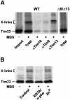
Fig. 2. Interaction of Tim8 and Tim13 with translocation intermediates of Tim23. (A) WT mitochondria were incubated with Tim23 precursor for 15 min at 25°C in the absence of Δψ (Import). Then 100 µM MBS was added and samples were incubated on ice for 30 min (Total). Aliquots were removed, mitochondria were lysed with 1% SDS and the samples were then diluted 20-fold with Tris-buffered saline containing 0.5% Triton X-100. After a clarifying spin the supernatant fractions were subjected to immunoprecipitation with antibodies against Tim10, Tim8 and Tim13. Right panel, cross-linking of Tim23 in Δ8/Δ13 mitochondria. X-links, Tim23-specific adducts. (B) Zn2+-dependent interaction of Tim8 and Tim13 with Tim23 precursor. WT mitochondria (Control) were incubated with EDTA/o-phe, and, where indicated, re-isolated in the presence of 0.1 mM ZnCl2 (see above). Subsequently Tim23 precursor was imported in the absence of Δψ and the samples were cross-linked with MBS.
None
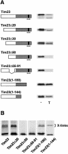
Fig. 3. Interaction of the TIM8–13 complex with the N-terminal portion of Tim23. (A) Translocation of Tim23-derived precursors across the outer membrane. Tim23-derived precursor constructs are schematically outlined. The grey box indicates the C-terminal portion, which is integrated into the inner membrane, the black box and the +++ refer to the Δψ-dependent import signal (Káldi et al., 1998). Numbers refer to amino acid residues of Tim23, deletions are indicated by Δ. The precursors were incubated with WT mitochondria in the absence of Δψ for 15 min at 25°C. Aliquots were treated with100 µg/ml trypsin (T). (B) Cross-linking of Tim8 and Tim13 with Tim23-derived precursor constructs. Precursors were imported into WT mitochondria in the absence of Δψ and then cross-linked with 100 µM MBS.
None
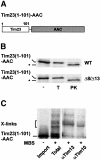
Fig. 4. Interaction of Tim23(1–101)–AAC with small Tim proteins of the intermembrane space. (A) Schematic outline of Tim23(1–101)–AAC. (B) Radiolabelled Tim23(1–101)–AAC precursor was imported for 10 min at 25°C into WT and Δ8/Δ13 mitochondria, which were energized with NADH. Samples were treated with100 µg/ml trypsin (T) or 200 µg/ml PK. *, fragment generated by cleavage of Tim23 by PK. (C) Cross-linking of Tim23(1–101)–AAC with small Tim proteins. Tim23(1–101)–AAC was imported into WT mitochondria in the absence of Δψ (Import) and then cross-linked with 100 µM MBS (Total). Aliquots were subjected to immunoprecipitation with antibodies against Tim13 and Tim10 as described in Figure 2. X-links, Tim23(1–101)–AAC-specific adducts.
None
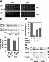
Fig. 5. The TIM8–13 complex facilitates import of Tim23 at low Δψ. (A) Cold-sensitive growth phenotype of Δ8/Δ13 cells in the presence of glucose. Δ8/Δ13 cells and the parental WT cells were grown at 30°C in YPD medium to an OD578 of 1. The cultures were subjected to serial 10-fold dilutions and 2 µl aliquots were spotted onto YPD and YPG agar plates. The plates were incubated for 2 days at 30°C and for 3 days at 15°C. (B) Assessment of Δψ. Mitochondria from WT cells (100 µg/ml) were incubated for 10 min in import buffer with 2 mM 3,3′-dipropylthiadicarbocyanide iodide. Where indicated the samples contained 8 µM antimycin, 20 µM oligomycin and 2.5 mM ATP to lower Δψ or 1 µM valinomycin (val) and 80 mM KCl to dissipateΔψ. Subsequently the mitochondria were removed by centrifugation and the fluorescence (excitation at 622 nm, emission at 670 nm) ofthe supernatant fractions was determined at 25°C (Rassow, 1999). (C) Import of Tim23 at low Δψ. WT and Δ8/Δ13 mitochondria were incubated with antimycin, oligomycin and ATP to generate a low Δψ. Tim23 precursor was imported for 15 min at 25 and 15°C and samples were treated with 200 µg of PK as indicated. Samples were analysed by SDS–PAGE (upper panel) and quantified with a phosphoimager (lower panel). (D) Import of precursors into mitoplasts at high and low Δψ. WT mitochondria were converted into mitoplasts by swelling in 20 mM HEPES–KOH pH 7.2. The mitoplasts were incubated with NADH or anti/oli. pSu9(1–94)-DHFR (upper panel) and Tim23 precursor (lower panel) were imported into the mitoplasts for 15 min at 25°C. When indicated the mitoplasts were treated with 200 µg/ml PK. p, precursor; m, mature form of Su9(1–94)-DHFR; Tim23′′, PK-resistant fragment of inserted Tim23.
None
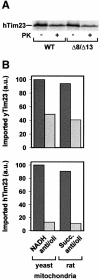
Fig. 6. Import of human Tim23. (A) Radiolabelled human Tim23 (hTim23) precursor was synthesized in reticulocyte lysate and imported for 15 min at 25°C into WT and Δ8/Δ13 mitochondria from yeast that were energized with NADH. Samples were treated with 100 µg/ml PK. (B) Import of human Tim23 requires high Δψ. The precursors of yeast Tim23 (yTim23) (upper panel) and human Tim23 (lower panel) were imported into yeast mitochondria and mitochondria from rat liver that were energized with NADH or succinate (Succ.), respectively. Samples were treated with 100 µg/ml PK, subjected to SDS–PAGE and imported Tim23 was quantified with a phosphoimaging system.
None
Fig. 7. Proposed role of the TIM8–13 complex in the import pathway of Tim23. Cytosolic Tim23 precursor (stage I) binds to receptors on the surface of the outer membrane (stage II). It is then translocated across the TOM complex and trapped by the TIM8–13 complex in the intermembrane space (stage IIIa). The TIM22 complex interacts with the accumulated precursor (stage IIIb) and mediates in a Δψ-dependent manner its release from the TOM complex and insertion into the inner membrane (stage IV). IM, inner membrane; OM, outer membrane.

Similar articles

Cited by

References

    1. Adam A., Endres,M., Sirrenberg,C., Lottspeich,F., Neupert,W. and Brunner,M. (1999) Tim9, a new component of the TIM22.54 translocase in mitochondria. EMBO J., 18, 313–319. - PMC - PubMed
    1. Bauer M.F., Sirrenberg,C., Neupert,W. and Brunner,M. (1996) Role of Tim23 as voltage sensor and presequence receptor in protein import into mitochondria. Cell, 87, 33–41. - PubMed
    1. Bauer M.F., Gempel,K., Reichert,A.S., Rappold,G.A., Lichtner,P., Gerbitz,K.-D., Neupert,W., Brunner,M. and Hofmann,S. (1999a) Genetic and structural characterization of the human mitochondrial inner membrane translocase. J. Mol. Biol., 289, 69–82. - PubMed
    1. Bauer M.F., Rothbauer,U., Mühlenbein,N., Smith,R.J.H., Gerbitz,K.-D., Neupert,W., Brunner,M. and Hofmann,S. (1999b) The mitochondrial TIM22 preprotein translocase is highly conserved throughout the eukaryotic kingdom. FEBS Lett., 464, 41–47. - PubMed
    1. Bauer M.F., Hofmann,S., Neupert,W. and Brunner,M. (2000) Protein translocation into mitochondria: the role of TIM complexes. Trends Cell Biol., 10, 25–31. - PubMed

Publication types

MeSH terms