

Spatial control of cell differentiation in Myxococcus xanthus
- PMID: 10922065
- PMCID: PMC16828
- DOI: 10.1073/pnas.97.16.9098
Spatial control of cell differentiation in Myxococcus xanthus
Abstract
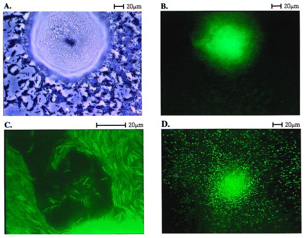
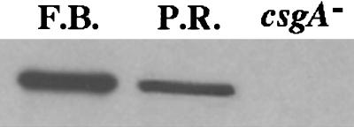
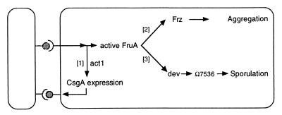
Myxococcus xanthus develops species-specific multicellular fruiting bodies. Starting from a uniform mat of cells, some cells enter into nascent fruiting body aggregates, whereas other cells remain outside. The cells within the fruiting body differentiate from rods into spherical, heat-resistant spores, whereas the cells outside the aggregates, called peripheral cells, remain rod-shaped. Early developmentally regulated genes are expressed in peripheral cells as well as by cells in the fruiting bodies. By contrast, late developmental genes are only expressed by cells within the nascent fruiting bodies. The data show that peripheral cells begin to develop, but are unable to express genes that are switched on later than about 6 h after the start of development. All of the genes whose expression is limited to the fruiting body are dependent on C-signaling either directly or indirectly, whereas the genes that are equally expressed in peripheral rods and in fruiting body cells are not. One of the C-signal-dependent and spatially patterned operons is called dev, and the dev operon has been implicated in the process of sporulation. It is proposed that expression of certain genes, including those of the dev operon, is limited to the nascent fruiting body because fruiting body cells engage in a high level of C-signaling. Peripheral cells do less C-signaling than fruiting body cells, because they have a different spatial arrangement and are at lower density. As a consequence, peripheral cells fail to express the late genes necessary for spore differentiation.
Figures
Similar articles
-
Two-Component Signal Transduction Systems That Regulate the Temporal and Spatial Expression of Myxococcus xanthus Sporulation Genes.J Bacteriol. 2015 Sep 14;198(3):377-85. doi: 10.1128/JB.00474-15. Print 2016 Feb 1. J Bacteriol. 2015. PMID: 26369581 Free PMC article. Review.
-
Global transcriptome analysis of spore formation in Myxococcus xanthus reveals a locus necessary for cell differentiation.BMC Genomics. 2010 Apr 26;11:264. doi: 10.1186/1471-2164-11-264. BMC Genomics. 2010. PMID: 20420673 Free PMC article.
-
The enhancer binding protein Nla6 regulates developmental genes that are important for Myxococcus xanthus sporulation.J Bacteriol. 2015 Apr;197(7):1276-87. doi: 10.1128/JB.02408-14. Epub 2015 Feb 2. J Bacteriol. 2015. PMID: 25645554 Free PMC article.
-
The dev Operon Regulates the Timing of Sporulation during Myxococcus xanthus Development.J Bacteriol. 2017 Apr 25;199(10):e00788-16. doi: 10.1128/JB.00788-16. Print 2017 May 15. J Bacteriol. 2017. PMID: 28264995 Free PMC article.
-
Coupling gene expression and multicellular morphogenesis during fruiting body formation in Myxococcus xanthus.Mol Microbiol. 2003 Apr;48(1):1-8. doi: 10.1046/j.1365-2958.2003.03399.x. Mol Microbiol. 2003. PMID: 12657040 Review.
Cited by
-
Structural basis for regulation of a CBASS-CRISPR-Cas defense island by a transmembrane anti-σ factor and its ECF σ partner.Sci Adv. 2024 Oct 25;10(43):eadp1053. doi: 10.1126/sciadv.adp1053. Epub 2024 Oct 25. Sci Adv. 2024. PMID: 39454004 Free PMC article.
-
The Myxococcus xanthus spore cuticula protein C is a fragment of FibA, an extracellular metalloprotease produced exclusively in aggregated cells.PLoS One. 2011;6(12):e28968. doi: 10.1371/journal.pone.0028968. Epub 2011 Dec 12. PLoS One. 2011. PMID: 22174937 Free PMC article.
-
The Regulation of LexA on UV-Induced SOS Response in Myxococcus xanthus Based on Transcriptome Analysis.J Microbiol Biotechnol. 2021 Jul 28;31(7):912-920. doi: 10.4014/jmb.2103.03047. J Microbiol Biotechnol. 2021. PMID: 34024894 Free PMC article.
-
Benzylsuccinate synthase of Azoarcus sp. strain T: cloning, sequencing, transcriptional organization, and its role in anaerobic toluene and m-xylene mineralization.J Bacteriol. 2001 Dec;183(23):6763-70. doi: 10.1128/JB.183.23.6763-6770.2001. J Bacteriol. 2001. PMID: 11698363 Free PMC article.
-
Four unusual two-component signal transduction homologs, RedC to RedF, are necessary for timely development in Myxococcus xanthus.J Bacteriol. 2005 Dec;187(23):8191-5. doi: 10.1128/JB.187.23.8191-8195.2005. J Bacteriol. 2005. PMID: 16291693 Free PMC article.
References
-
- Dworkin M, Gibson S. Science. 1964;146:243–244. - PubMed
Publication types
MeSH terms
Grants and funding
LinkOut - more resources
Full Text Sources
Research Materials
