. 2000 Aug;20(16):6138-46.
doi: 10.1128/MCB.20.16.6138-6146.2000.

Sp110 localizes to the PML-Sp100 nuclear body and may function as a nuclear hormone receptor transcriptional coactivator

Affiliations

Sp110 localizes to the PML-Sp100 nuclear body and may function as a nuclear hormone receptor transcriptional coactivator

D B Bloch et al. Mol Cell Biol. 2000 Aug.

Abstract

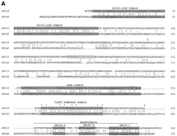
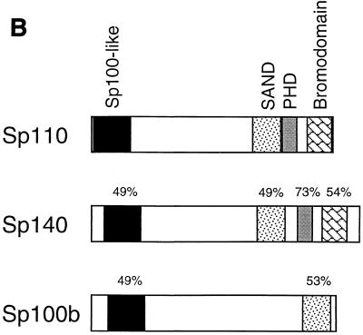
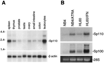
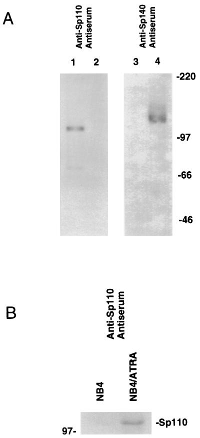
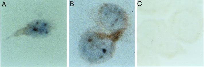
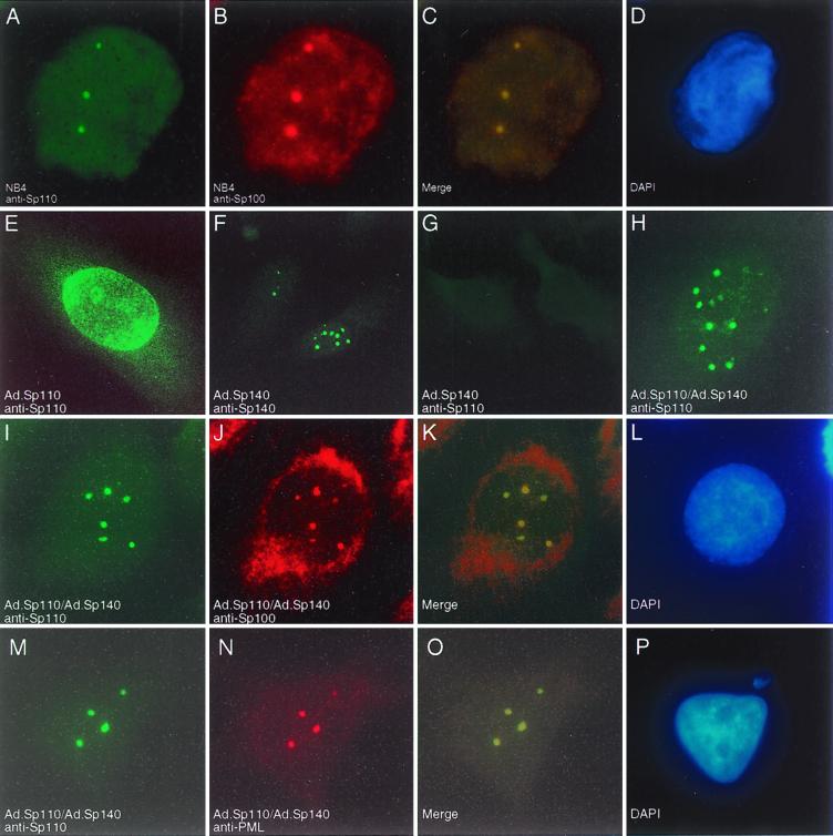
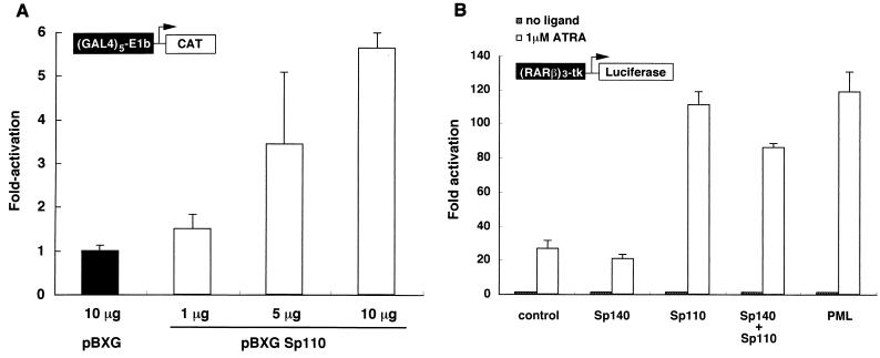
The nuclear body is a multiprotein complex that may have a role in the regulation of gene transcription. This structure is disrupted in a variety of human disorders including acute promyelocytic leukemia and viral infections, suggesting that alterations in the nuclear body may have an important role in the pathogenesis of these diseases. In this study, we identified a cDNA encoding a leukocyte-specific nuclear body component designated Sp110. The N-terminal portion of Sp110 was homologous to two previously characterized components of the nuclear body (Sp100 and Sp140). The C-terminal region of Sp110 was homologous to the transcription intermediary factor 1 (TIF1) family of proteins. High levels of Sp110 mRNA were detected in human peripheral blood leukocytes and spleen but not in other tissues. The levels of Sp110 mRNA and protein in the human promyelocytic leukemia cell line NB4 increased following treatment with all-trans retinoic acid (ATRA), and Sp110 localized to PML-Sp100 nuclear bodies in ATRA-treated NB4 cells. Because of the structural similarities between Sp110 and TIF1 proteins, the effect of Sp110 on gene transcription was examined. An Sp110 DNA-binding domain fusion protein activated transcription of a reporter gene in transfected mammalian cells. In addition, Sp110 produced a marked increase in ATRA-mediated expression of a reporter gene containing a retinoic acid response element. Taken together, the results of this study demonstrate that Sp110 is a member of the Sp100/Sp140 family of nuclear body components and that Sp110 may function as a nuclear hormone receptor transcriptional coactivator. The predominant expression of Sp110 in leukocytes and the enhanced expression of Sp110 in NB4 cells treated with ATRA raise the possibility that Sp110 has a role in inducing differentiation of myeloid cells.


Figures

FIG. 1
FIG. 1
(A) The predicted amino acid sequence of Sp110 and comparison with Sp140. Sp110 has a modular structure that includes the “Sp100-like domain,” SAND domain, plant homeobox domain, and bromodomain (shaded). Dashed box, presumed nuclear localization sequence in Sp110, between amino acids 288 and 306; asterisks, conserved cysteine and histidine residues in the plant homeobox domain; solid box, LXXLL-type nuclear hormone receptor interaction domain in Sp110. The amino acid sequence of the IFN-inducible protein nuclear phosphoprotein 72 is contained within the sequence of Sp110 beginning at methionine 241 and extending to leucine 605 (arrows). (B) Amino acid sequence homology between Sp110 and Sp140 and between Sp110 and Sp100b. Regions of homology in the Sp100-like region, SAND domain, plant homeobox domain, and bromodomain are indicated.
FIG. 1
FIG. 1
(A) The predicted amino acid sequence of Sp110 and comparison with Sp140. Sp110 has a modular structure that includes the “Sp100-like domain,” SAND domain, plant homeobox domain, and bromodomain (shaded). Dashed box, presumed nuclear localization sequence in Sp110, between amino acids 288 and 306; asterisks, conserved cysteine and histidine residues in the plant homeobox domain; solid box, LXXLL-type nuclear hormone receptor interaction domain in Sp110. The amino acid sequence of the IFN-inducible protein nuclear phosphoprotein 72 is contained within the sequence of Sp110 beginning at methionine 241 and extending to leucine 605 (arrows). (B) Amino acid sequence homology between Sp110 and Sp140 and between Sp110 and Sp100b. Regions of homology in the Sp100-like region, SAND domain, plant homeobox domain, and bromodomain are indicated.
FIG. 2
FIG. 2
(A) Identification of Sp110 mRNA in human tissues by RNA blot hybridization. A membrane containing 2.5 μg of poly(A)+ RNA per lane from human spleen, thymus, prostate, testis, ovary, small intestine, colon, and peripheral blood leukocytes was hybridized with a 32P-radiolabeled XbaI restriction fragment of the Sp110 cDNA. After being washed under stringent conditions, the membrane was exposed to autoradiography. High levels of mRNA encoding Sp110 were detected in human spleen and peripheral blood leukocytes. To confirm the presence of RNA in each lane, the membrane was subsequently hybridized with a radiolabeled human β-actin cDNA probe. (B) Expression of Sp110 mRNA in myeloid precursor cell lines. Low levels of Sp110 mRNA were detected in NB4 cells (lane NB4) and in HL60 cells (lane HL60). Treatment of NB4 cells with ATRA (1 μM) for 48 h induced expression of Sp110 (lane NB4/ATRA). Treatment of HL60 cells with IFN-γ (200 U/ml) for 48 h also markedly increased Sp110 gene expression (lane HL60/IFN). Similar changes in Sp100 mRNA were observed in ATRA-treated NB4 cells and IFN-γ-treated HL60 cells. Ethidium bromide staining of 28S RNA confirmed equal loading of RNA samples.
FIG. 3
FIG. 3
(A) Immunoblotting of adenovirus-infected HEp-2 cells using anti-Sp110 and anti-Sp140 antisera. Immunoblots were prepared from extracts of HEp-2 cells infected with Ad.Sp110 (lanes 1 and 3) or Ad.Sp140 (lanes 2 and 4). Anti-Sp110 antiserum reacted with Sp110 in Ad.Sp110-infected HEp-2 cells (lane 1) but not with Sp140 in Ad.Sp140-infected HEp-2 cells (lane 2) or Sp100 (normally expressed in HEp-2 cells). Anti-Sp140 antiserum reacted with Sp140 in Ad.Sp140-infected HEp-2 cells (lane 4) but not with Sp110 in Ad.Sp110-infected HEp-2 cells (lane 3). (B) Immunoblot of ATRA-treated and control NB4 cells. Immunoblots were prepared from extracts of NB4 cells treated for 48 h with ATRA (1 μM; lane NB4/ATRA) or control NB4 cells (lane NB4). ATRA treatment increased the level of immunoreactive Sp110.
FIG. 4
FIG. 4
Immunohistochemical localization of Sp110 in NB4 cells. Control NB4 cells and NB4 cells treated for 48 h with ATRA (1 μM) were subjected to cytospin centrifugation, fixed, and stained with rat anti-Sp110 antiserum. Staining was observed within dot-like regions in the nuclei of ATRA-treated NB4 cells (A and B). Anti-Sp110 antiserum did not react with untreated NB4 cells (C).
FIG. 5
FIG. 5
Immunofluorescence microscopy of ATRA-treated NB4 cells and adenovirus-infected HEp-2 cells. NB4 cells were treated for 48 h with ATRA (1 μM) and were stained with rat anti-Sp110 antiserum (A) and human serum containing anti-Sp100 antibodies (B). Sp110 colocalized with Sp100 in nuclear bodies in ATRA-treated NB4 cells (merging of green and red fluorescence and DAPI [4′,6′-diamidino-2-phenylindole] staining are shown in panels C and D, respectively). To further investigate the cellular location of Sp110, HEp-2 cells, which normally do not express either Sp110 or Sp140, were infected with Ad.Sp110 and stained with anti-Sp110 antiserum. Sp110 was observed in a granular pattern within the nucleus and appeared to associate with the nuclear membrane (E). In contrast, HEp-2 cells infected with Ad.Sp140 and stained with anti-Sp140 antiserum revealed a typical nuclear body staining pattern (F). No fluorescence was seen in cells infected with Ad.Sp140 and stained with anti-Sp110 antiserum (G), confirming the specificity of this antiserum for Sp110. In cells infected with both Ad.Sp110 and Ad.Sp140, Sp110 localized to nuclear bodies (H, I, and M). Staining of cells infected with both Ad.Sp110 and Ad.Sp140 with anti-Sp110 antiserum (I) and anti-Sp100 antibodies (J) revealed colocalization of the two proteins (K). In addition, staining of infected cells with anti-Sp110 (M) and anti-PML (N) antibodies demonstrated colocalization of the two proteins (O). (L and P), DAPI staining.
FIG. 6
FIG. 6
(A) Sp110 acts as a transcriptional activator when tethered to DNA. Plasmids encoding the GAL4 DNA-binding domain fused to Sp110 (pBXG-Sp110) or the GAL4 DNA-binding domain alone were transfected into COS cells together with a reporter plasmid directing expression of CAT under the control of a GAL4 DNA-binding domain response element. A plasmid encoding growth hormone was included as a control for efficiency of transfection, and transfections were performed in triplicate. The total amount of plasmid DNA was the same in each transfection. Results are means ± standard errors of the means (SEM). CAT activity in pBXG-Sp110-transfected cells was expressed as the fold increase compared with the activity in pBXG-transfected cells. Production of growth hormone in cells transfected with pBXG did not differ from that in cells transfected with pBXG-Sp110. Transfection of COS cells with 1, 5, and 10 μg of pBXG-Sp110 increased CAT activity in a DNA dose-dependent manner. (B) Sp110 may function as a nuclear hormone receptor transcriptional coactivator. Plasmids encoding Sp110, Sp140, Sp110 and Sp140, or PML or vector alone was transfected into COS cells together with a reporter plasmid containing the luciferase gene driven by three copies of the RARα response element derived from the RARβ promoter region. Results are fold increases in luciferase activity in ATRA-treated versus untreated transfected cells. Transfections were performed in triplicate, and results are presented as means ± SEM. The results are representative of three separate experiments. Expression of Sp110 enhanced ATRA-induced responsiveness compared with vector alone. The extent of enhanced ATRA responsiveness by Sp110 was similar to that induced by PML. In contrast, Sp140 did not increase ATRA-induced expression of the reporter gene and expression of both Sp140 and Sp110 did not enhance luciferase activity to a greater extent than did expression of Sp110 alone.

Similar articles

Cited by

References

    1. Aasland R, Gibson T J, Stewart A F. The PHD finger: implications for chromatin-mediated transcriptional regulation. Trends Biochem Sci. 1995;20:56–59. - PubMed
    1. Alam J, Cook J L. Reporter genes: application to the study of mammalian gene transcription. Anal Biochem. 1990;188:245–254. - PubMed
    1. Ascoli C A, Maul G G. Identification of a novel nuclear domain. J Cell Biol. 1991;112:785–795. - PMC - PubMed
    1. Bloch D B, Chiche J-D, Orth D, Rosenzweig A, Bloch K D. Structural and functional hetereogeneity of nuclear bodies. Mol Cell Biol. 1999;19:4423–4430. - PMC - PubMed
    1. Bloch D B, de la Monte S M, Guigaouri P, Filippov A, Bloch K D. Identification and characterization of a leukocyte-specific component of the nuclear body. J Biol Chem. 1996;46:29198–29204. - PubMed

Publication types

MeSH terms