. 2000 May 29;149(5):1039-52.
doi: 10.1083/jcb.149.5.1039.

PNG1, a yeast gene encoding a highly conserved peptide:N-glycanase

Affiliations

PNG1, a yeast gene encoding a highly conserved peptide:N-glycanase

T Suzuki et al. J Cell Biol. .

Abstract

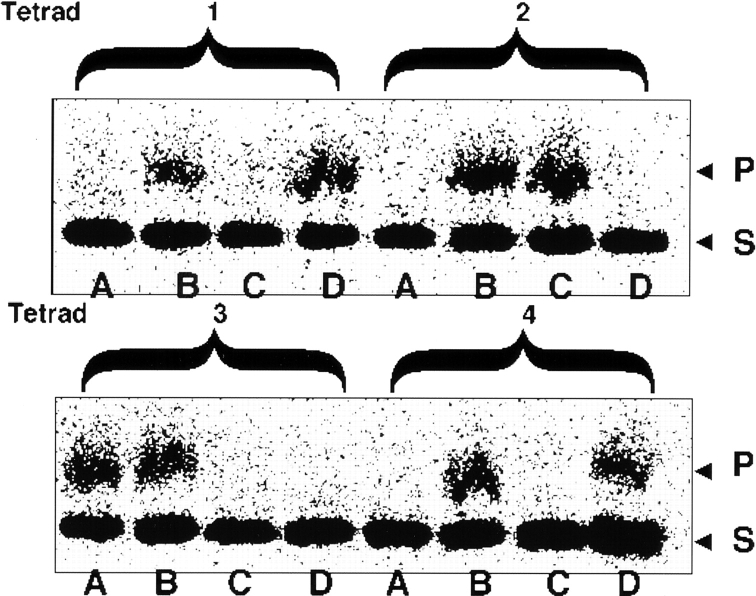
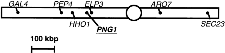
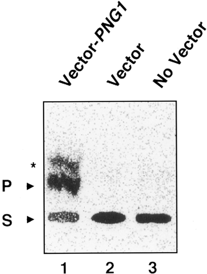
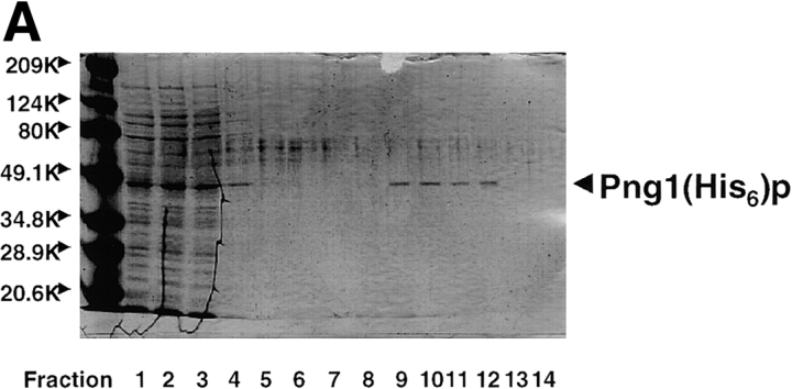
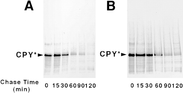
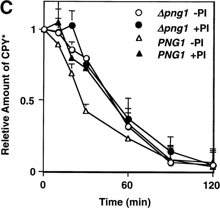
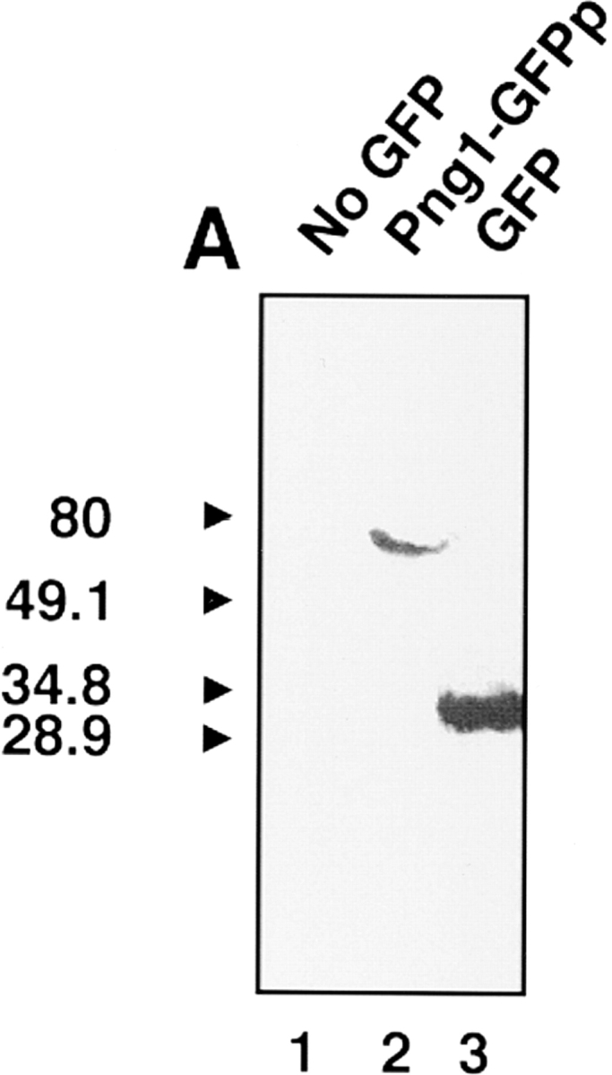
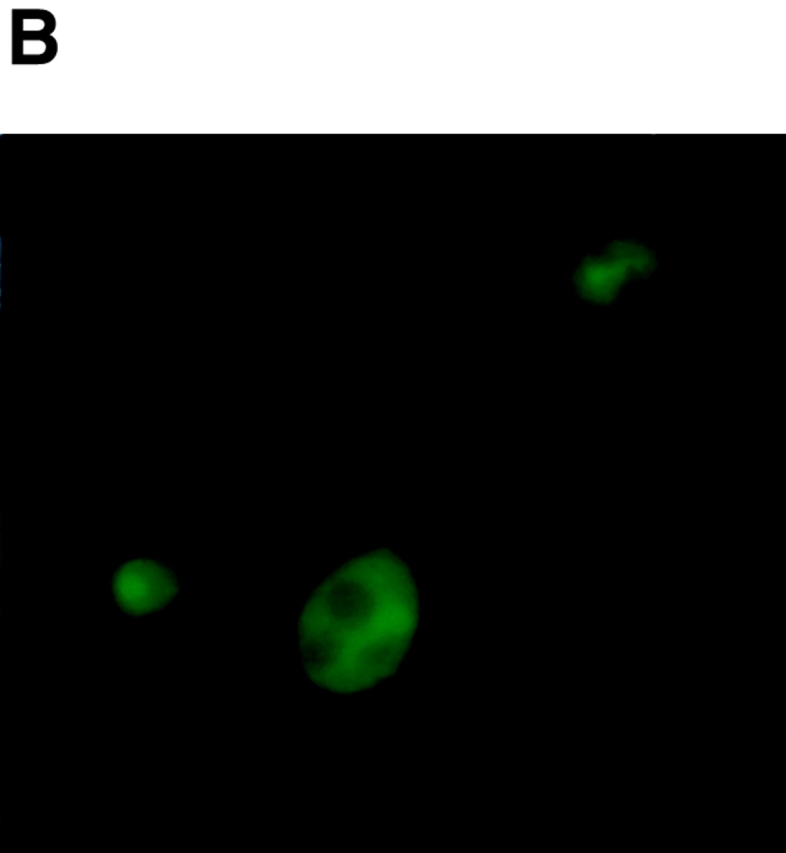
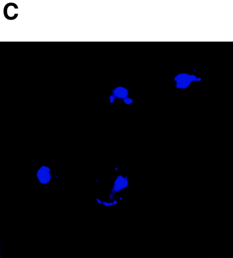
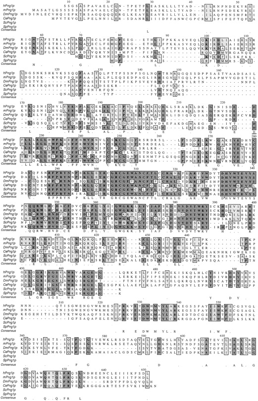
It has been proposed that cytoplasmic peptide:N-glycanase (PNGase) may be involved in the proteasome-dependent quality control machinery used to degrade newly synthesized glycoproteins that do not correctly fold in the ER. However, a lack of information about the structure of the enzyme has limited our ability to obtain insight into its precise biological function. A PNGase-defective mutant (png1-1) was identified by screening a collection of mutagenized strains for the absence of PNGase activity in cell extracts. The PNG1 gene was mapped to the left arm of chromosome XVI by genetic approaches and its open reading frame was identified. PNG1 encodes a soluble protein that, when expressed in Escherichia coli, exhibited PNGase activity. PNG1 may be required for efficient proteasome-mediated degradation of a misfolded glycoprotein. Subcellular localization studies indicate that Png1p is present in the nucleus as well as the cytosol. Sequencing of expressed sequence tag clones revealed that Png1p is highly conserved in a wide variety of eukaryotes including mammals, suggesting that the enzyme has an important function.


Figures

Figure 1
Figure 1
Assay of PNGase activity in four sets of tetrads from the cross of a PNGase-defective mutant (No. 352) with a wild-type strain (PS593). Protein extracts (∼20 μg) from each spore colony were incubated with 25 μM of [14C]asialofetuin peptide I in 6 μl of 70 mM Hepes-NaOH buffer, pH 7.2, and 5 mM DTT at 25°C for 16 h. The reaction product was analyzed by paper chromatography and the radioactive peptides were visualized using a PhosphorImager. A paper chromatogram of four different tetrads (1–4) are shown. P, de-N-glycosylated product ([14C]-Leu-Asp-Asn-Ser-Arg); and S, substrate ([14C]-Leu-Asn(GlcNAc5 Man3Gal3)-Asn-Ser-Arg).
Figure 2
Figure 2
Schematic representation of chromosome XVI of S. cerevisiae and the marker genes used in this study. The PNG1 locus determined in this study is also indicated.
Figure 3
Figure 3
Assay of PNGase activity in an E. coli cell-free extract. The E. coli strain used was BL21(DE3)pLysS. Shown is a paper chromatogram of the reaction product formed after 10 min of incubation of the E. coli extract with labeled substrate. (lane 1) E. coli extract with pET-28b-PNG1; (lane 2) E. coli extract with pET-28b (control vector); and (lane 3) E. coli extract without vector. The extra minor band indicated with an asterisk was confirmed to be [14C]Leu-Asp by analysis by paper electrophoresis, as described earlier (Kitajima, et al. 1995), and may be derived from a contaminating protease activity in the E. coli extract. In fact, this degradation product represents a fraction of the product of PNGase activity because the second amino acid in the substrate was converted into Asp instead of remaining as Asn. P, de-N-glycosylated product ([14C]-Leu-Asp-Asn-Ser-Arg); and S, substrate ([14C]-Leu-Asn(Glc NAc5Man3Gal3)-Asn-Ser-Arg). For details, see Materials and Methods.
Figure 4
Figure 4
Purification of (His6)-tagged Png1p (Png1(His6)p) and reaction product analysis of [14C]asialofetuin glycopeptide I with it. E. coli extract overexpressing Png1(His6)p was incubated with TALON metal affinity resin, transferred to a 3-ml gravity flow column, and the elution was carried out as described in Materials and Methods. (A) Elution profile of each fraction was analyzed by 10% SDS-PAGE. 10 μl of each fraction was applied for the analysis. The position of migration of Png1(His6)p is indicated. Molecular masses indicated are based on prestained molecular mass marker standards (Bio-Rad). (B) Detection of PNGase activity in each elution fraction. The activity in each fraction was measured after 16-h incubation of reaction mixture at room temperature. (C and D) Analysis of reaction products produced by incubation of [14C]asialofetuin glycopeptide I with purified Png1(His6)p using (C) paper chromatography and (D) paper electrophoresis. (lane 1) [14C]asialofetuin glycopeptide I; (lane 2) reaction product formed by Png1(His6)p; and (lane 3) reference reaction product formed by PNGase F. The PNGase-deglycosylated product, [14C]-Leu-Asp-Asp-Ser-Arg, is indicated by asterisks.
Figure 4
Figure 4
Purification of (His6)-tagged Png1p (Png1(His6)p) and reaction product analysis of [14C]asialofetuin glycopeptide I with it. E. coli extract overexpressing Png1(His6)p was incubated with TALON metal affinity resin, transferred to a 3-ml gravity flow column, and the elution was carried out as described in Materials and Methods. (A) Elution profile of each fraction was analyzed by 10% SDS-PAGE. 10 μl of each fraction was applied for the analysis. The position of migration of Png1(His6)p is indicated. Molecular masses indicated are based on prestained molecular mass marker standards (Bio-Rad). (B) Detection of PNGase activity in each elution fraction. The activity in each fraction was measured after 16-h incubation of reaction mixture at room temperature. (C and D) Analysis of reaction products produced by incubation of [14C]asialofetuin glycopeptide I with purified Png1(His6)p using (C) paper chromatography and (D) paper electrophoresis. (lane 1) [14C]asialofetuin glycopeptide I; (lane 2) reaction product formed by Png1(His6)p; and (lane 3) reference reaction product formed by PNGase F. The PNGase-deglycosylated product, [14C]-Leu-Asp-Asp-Ser-Arg, is indicated by asterisks.
Figure 5
Figure 5
Complementation of the defect of PNGase activity in png1Δ cells by plasmids bearing PNG1. (A) PNGase activity assay in extract of TSY146 (png1Δ) cells with pRS316 (negative control; lane 1), pRS316-PNG1 (CEN plasmid bearing PNG1; lane 2) and YEp352-PNG1 (2-micron plasmid bearing PNG1). (B) Time course study of PNGase activity in TSY146 cells bearing pRS316-PNG1 (CEN, x) or YEp352-PNG1 (2 microns; •). At the indicated times, an aliquot of reaction mixture was removed and PNGase activity was quantitated using paper chromatography.
Figure 5
Figure 5
Complementation of the defect of PNGase activity in png1Δ cells by plasmids bearing PNG1. (A) PNGase activity assay in extract of TSY146 (png1Δ) cells with pRS316 (negative control; lane 1), pRS316-PNG1 (CEN plasmid bearing PNG1; lane 2) and YEp352-PNG1 (2-micron plasmid bearing PNG1). (B) Time course study of PNGase activity in TSY146 cells bearing pRS316-PNG1 (CEN, x) or YEp352-PNG1 (2 microns; •). At the indicated times, an aliquot of reaction mixture was removed and PNGase activity was quantitated using paper chromatography.
Figure 6
Figure 6
Effect of png1Δ on CPY* degradation. Radiolabeled CPY*, prepared as described in Materials and Methods, was analyzed by 7.5% SDS-PAGE, and the labeled protein was visualized using a PhosphorImager. (A) CPY* from TSY147 (BY4742 prc1-1); and (B) CPY* from TSY149 (BY4742 prc1-1 png1Δ::KanMX4). (C) Effect of proteasome inhibitor, MG-132, on the degradation of CPY*. The CPY* recovered by immunoprecipitation at the times indicated was quantitated. PI, proteasome inhibitor (MG-132). The values are the average of three separate experiments and standard errors are indicated. For details, see Materials and Methods.
Figure 6
Figure 6
Effect of png1Δ on CPY* degradation. Radiolabeled CPY*, prepared as described in Materials and Methods, was analyzed by 7.5% SDS-PAGE, and the labeled protein was visualized using a PhosphorImager. (A) CPY* from TSY147 (BY4742 prc1-1); and (B) CPY* from TSY149 (BY4742 prc1-1 png1Δ::KanMX4). (C) Effect of proteasome inhibitor, MG-132, on the degradation of CPY*. The CPY* recovered by immunoprecipitation at the times indicated was quantitated. PI, proteasome inhibitor (MG-132). The values are the average of three separate experiments and standard errors are indicated. For details, see Materials and Methods.
Figure 7
Figure 7
Western blot analysis and subcellular localization of Png1p-GFP fusion protein (Png1-GFPp). (A) Western blotting of Png1-GFPp using anti-GFP antibody. (lane 1) TSY146 cells with pRS316 (CEN URA3; Sikorski and Hieter 1989) as a negative control; (lane 2) TSY146 with pPNG1-GFP (URA3); and (lane 3) TSY146 with pGFP-C-FUS (Niedenthal et al., 1996) as a positive control of GFP protein. (B) Localization of Png1-GFPp in S. cerevisiae. (C) DAPI staining of the same image shown in B.
Figure 7
Figure 7
Western blot analysis and subcellular localization of Png1p-GFP fusion protein (Png1-GFPp). (A) Western blotting of Png1-GFPp using anti-GFP antibody. (lane 1) TSY146 cells with pRS316 (CEN URA3; Sikorski and Hieter 1989) as a negative control; (lane 2) TSY146 with pPNG1-GFP (URA3); and (lane 3) TSY146 with pGFP-C-FUS (Niedenthal et al., 1996) as a positive control of GFP protein. (B) Localization of Png1-GFPp in S. cerevisiae. (C) DAPI staining of the same image shown in B.
Figure 8
Figure 8
Comparison of sequences of Png1p and its homologues. ScPng1p, S. cerevisiae Png1p; hPng1p, human Png1p homologue (partial; sequence was determined from EST clone 97076 [ATCC] and IMAGE clone ID 1316890); mPng1p, mouse Png1p homologue (sequence determined from IMAGE clone 948982); DmPng1p, D. melanogaster Png1p homologue (sequence determined from IMAGE clone LD46390); CePng1p, C. elegans Png1p homologue (sequence determined from yk491h3 and the Entrez database CAB57916); and SpPng1p, S. pombe Png1p homologue (sequence obtained from the Entrez database CAA21253). Asterisks represent the conserved cysteine residues in CXYC motifs. The conserved His residue that was found to be mutated to Tyr in the png1-1 allele is also indicated (#). Numbering is based on the mPng1p sequence.

Similar articles

Cited by

References

    1. Bebök Z., Mazzochi C., King S.A., Hong J.S., Sorscher E.J. The mechanism underlying cystic fibrosis transmembrane conductance regulator transport from the endoplasmic reticulum to the proteasome includes Sec61β and a cytosolic, deglycosylated intermediary. J. Biol. Chem. 1998;273:29873–29878. - PubMed
    1. Berger S., Menudier A., Julien R., Karamanos Y. Do de-N-glycosylation enzymes have an important role in plant cells? Biochimie. 1995;77:751–760. - PubMed
    1. Brodsky J.L., McCracken A.A. ER protein quality control and proteasome-mediated protein degradation. Semin. Cell Dev. Biol. 1999;10:507–513. - PubMed
    1. Cresswell P., Hughes E.A. Protein degradationthe ins and outs of the matter. Curr. Biol. 1997;7:R552–R555. - PubMed
    1. de Virgilio M., Weninger H., Ivessa N.E. Ubiquitination is required for the retro-translocation of a short-lived luminal endoplasmic reticulum glycoprotein to the cytosol for degradation by the proteasome. J. Biol. Chem. 1998;273:9734–9743. - PubMed

Publication types

MeSH terms

Substances