

Polycystin 1 is required for the structural integrity of blood vessels
- PMID: 10677526
- PMCID: PMC26504
- DOI: 10.1073/pnas.040550097
Polycystin 1 is required for the structural integrity of blood vessels
Abstract
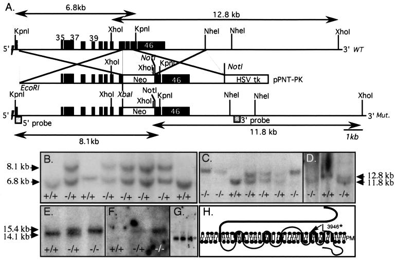
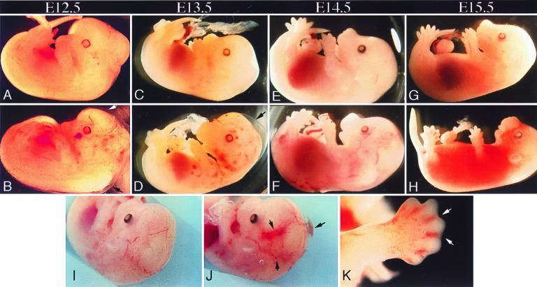
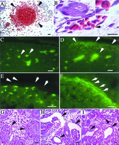
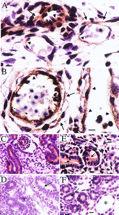
Autosomal dominant polycystic kidney disease (ADPKD), often caused by mutations in the PKD1 gene, is associated with life-threatening vascular abnormalities that are commonly attributed to the frequent occurrence of hypertension. A previously reported targeted mutation of the mouse homologue of PKD1 was not associated with vascular fragility, leading to the suggestion that the vascular lesion may be of a secondary nature. Here we demonstrate a primary role of PKD1 mutations in vascular fragility. Mouse embryos homozygous for the mutant allele (Pkd1(L)) exhibit s.c. edema, vascular leaks, and rupture of blood vessels, culminating in embryonic lethality at embryonic day 15.5. Kidney and pancreatic ductal cysts are present. The Pkd1-encoded protein, mouse polycystin 1, was detected in normal endothelium and the surrounding vascular smooth muscle cells. These data reveal a requisite role for polycystin 1 in maintaining the structural integrity of the vasculature as well as epithelium and suggest that the nature of the PKD1 mutation contributes to the phenotypic variance in ADPKD.
Figures
Similar articles
-
Perinatal lethality with kidney and pancreas defects in mice with a targetted Pkd1 mutation.Nat Genet. 1997 Oct;17(2):179-81. doi: 10.1038/ng1097-179. Nat Genet. 1997. PMID: 9326937
-
A human PKD1 transgene generates functional polycystin-1 in mice and is associated with a cystic phenotype.Hum Mol Genet. 2000 Nov 1;9(18):2617-27. doi: 10.1093/hmg/9.18.2617. Hum Mol Genet. 2000. PMID: 11063721
-
Comparison of Pkd1-targeted mutants reveals that loss of polycystin-1 causes cystogenesis and bone defects.Hum Mol Genet. 2001 Oct 1;10(21):2385-96. doi: 10.1093/hmg/10.21.2385. Hum Mol Genet. 2001. PMID: 11689485
-
Autosomal dominant polycystic kidney disease (ADPKD, MIM 173900, PKD1 and PKD2 genes, protein products known as polycystin-1 and polycystin-2).Eur J Hum Genet. 2004 May;12(5):347-54. doi: 10.1038/sj.ejhg.5201162. Eur J Hum Genet. 2004. PMID: 14872199 Review.
-
Molecular genetics and mechanism of autosomal dominant polycystic kidney disease.Mol Genet Metab. 2000 Jan;69(1):1-15. doi: 10.1006/mgme.1999.2943. Mol Genet Metab. 2000. PMID: 10655152 Review.
Cited by
-
Mouse models of polycystic kidney disease induced by defects of ciliary proteins.BMB Rep. 2013 Feb;46(2):73-9. doi: 10.5483/bmbrep.2013.46.2.022. BMB Rep. 2013. PMID: 23433108 Free PMC article. Review.
-
Mutation analysis of the entire PKD1 gene: genetic and diagnostic implications.Am J Hum Genet. 2001 Jan;68(1):46-63. doi: 10.1086/316939. Epub 2000 Dec 12. Am J Hum Genet. 2001. PMID: 11115377 Free PMC article.
-
The mechanosensory role of primary cilia in vascular hypertension.Int J Vasc Med. 2011;2011:376281. doi: 10.1155/2011/376281. Epub 2011 Jun 16. Int J Vasc Med. 2011. PMID: 21748021 Free PMC article.
-
Modulation of the secretory pathway rescues zebrafish polycystic kidney disease pathology.J Am Soc Nephrol. 2014 Aug;25(8):1749-59. doi: 10.1681/ASN.2013101060. Epub 2014 Mar 13. J Am Soc Nephrol. 2014. PMID: 24627348 Free PMC article.
-
Genetic variation in severe cystic fibrosis liver disease is associated with novel mechanisms for disease pathogenesis.Hepatology. 2024 Nov 1;80(5):1012-1025. doi: 10.1097/HEP.0000000000000863. Epub 2024 Mar 27. Hepatology. 2024. PMID: 38536042
References
-
- Dalgaard O Z. Acta Med Scand. 1957;328,Suppl.:1–255. - PubMed
-
- Grantham J J. Adv Intern Med. 1993;38:409–420. - PubMed
-
- Baert L. Kidney Int. 1978;13:519–525. - PubMed
-
- Gabow P A. N Eng J Med. 1993;329:332–342. - PubMed
-
- Huston J D, Torres V E, Sulivan P P, Offord K P, Wiebers D O. J Am Soc Nephrol. 1993;3:1871–1877. - PubMed
Publication types
MeSH terms
Substances
Grants and funding
LinkOut - more resources
Full Text Sources
Other Literature Sources
Molecular Biology Databases
