. 2000 Jan;11(1):325-37.
doi: 10.1091/mbc.11.1.325.

Identification of filamin as a novel ligand for caveolin-1: evidence for the organization of caveolin-1-associated membrane domains by the actin cytoskeleton

Affiliations
Free PMC article

Identification of filamin as a novel ligand for caveolin-1: evidence for the organization of caveolin-1-associated membrane domains by the actin cytoskeleton

M Stahlhut et al. Mol Biol Cell. 2000 Jan.
Free PMC article

Abstract

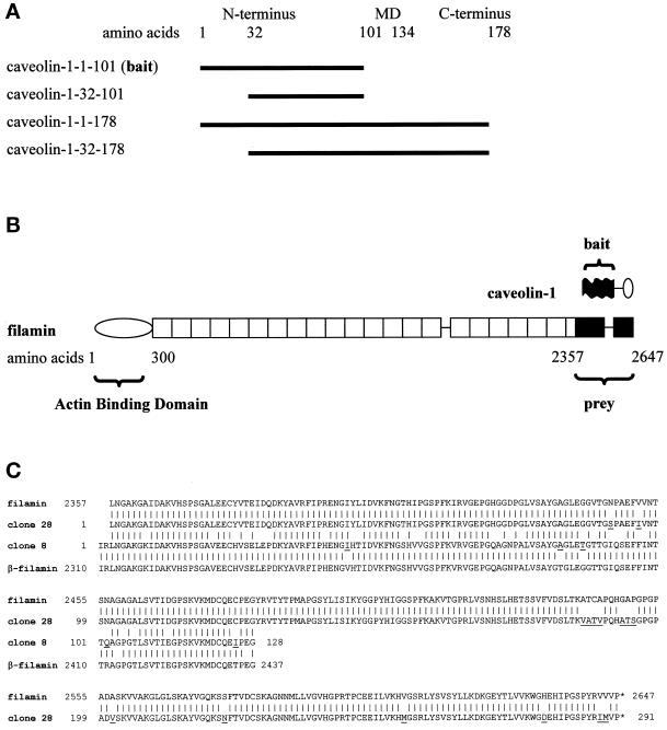
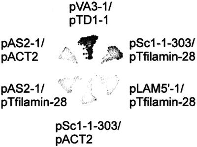
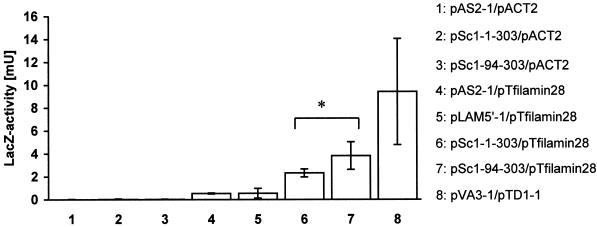
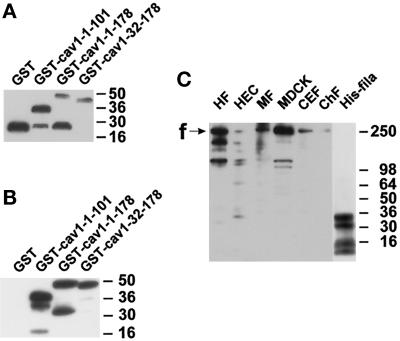
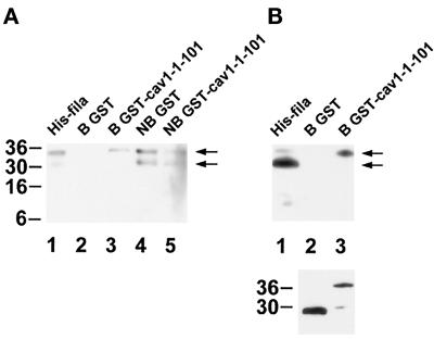
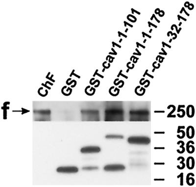
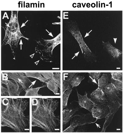
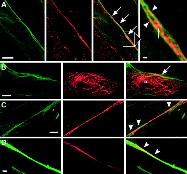
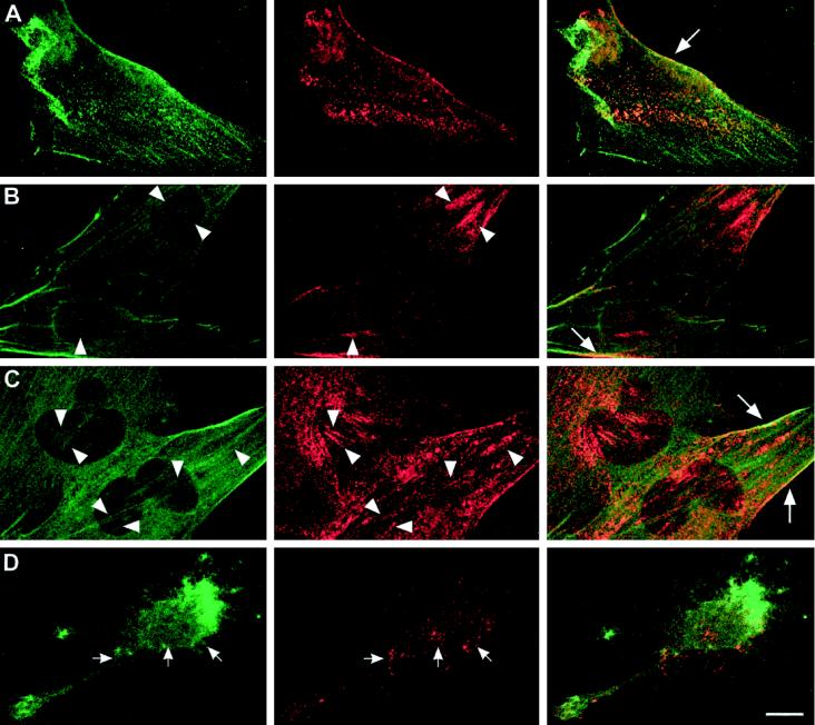
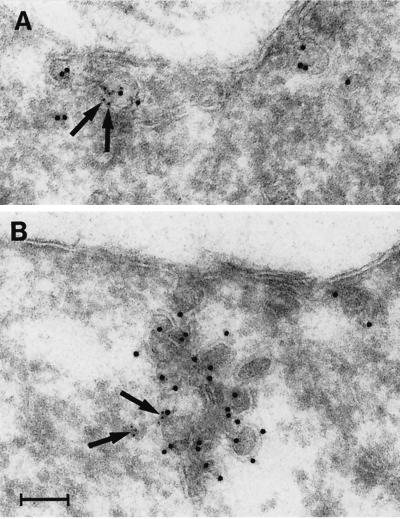
Reports on the ultrastructure of cells as well as biochemical data have, for several years, been indicating a connection between caveolae and the actin cytoskeleton. Here, using a yeast two-hybrid approach, we have identified the F-actin cross-linking protein filamin as a ligand for the caveolae-associated protein caveolin-1. Binding of caveolin-1 to filamin involved the N-terminal region of caveolin-1 and the C terminus of filamin close to the filamin-dimerization domain. In in vitro binding assays, recombinant caveolin-1 bound to both nonmuscle and muscle filamin, indicating that the interaction might not be cell type specific. With the use of confocal microscopy, colocalization of caveolin-1 and filamin was observed in elongated patches at the plasma membrane. Remarkably, when stress fiber formation was induced with Rho-stimulating Escherichia coli cytotoxic necrotizing factor 1, the caveolin-1-positive structures became coaligned with stress fibers, indicating that there was a physical link connecting them. Immunogold double-labeling electron microscopy confirmed that caveolin-1-labeled racemose caveolae clusters were positive for filamin. The actin network, therefore, seems to be directly involved in the spatial organization of caveolin-1-associated membrane domains.


Figures

Figure 1
Figure 1
(A) Domain organization of caveolin-1. Amino acids 1–101 represent the N-terminal domain, amino acids 102–134 represent the membrane domain (MD), and amino acids 135–178 represent the C-terminal domain. Two variants of the N-terminal domain (caveolin-1-1–101 and caveolin-1-32–101) and the full-length proteins caveolin-1α (caveolin-1-1–178) and caveolin-1β (caveolin-1-32–178) were analyzed. Caveolin-1-1–101 was used as the bait in the two-hybrid screen. (B) Domain organization of filamin and comparison of the sizes of caveolin-1 and filamin. (Top) The N terminus (black) of caveolin-1α was used as the bait in the two-hybrid screen. The membrane domain and the C-terminal domain of caveolin-1 are indicated by a line and an oval, respectively. (Bottom) The rod-like filamin molecule has an N-terminal actin-binding domain followed by 24 homologous repeats of 96 amino acids each. The fragment of filamin obtained in the two-hybrid screen (prey) is shown in black. Two hinges are present at the connections between repeats 15–16 and 23–24. (C) Alignment of the amino acid sequences of the human filamin and β-filamin isoforms and the two amino acid sequences of the filamin fragments isolated in the two-hybrid screening. Over the sequenced region, human filamin (GenBank accession number P21333) and human β-filamin (GenBank accession number AF042166) are 95 and 96% identical to clones 28 and 8, respectively. Clone 28 is 76% identical to clone 8. The high homologies clearly identified clones 28 and 8 as fragments of mouse filamin and β-filamin, respectively. Species-specific differences in the amino acid sequences in the two-hybrid clones are underlined.
Figure 2
Figure 2
Growth of Y190 clones on SD/−H/−W/−L minimal medium containing 25 mM 3-aminotriazole. Yeast strain Y190 was transformed with the indicated combinations of plasmids. Only yeast cells coexpressing caveolin-1-1–101 and filamin-28 (pSc1-1–303/pTfilamin-28) and p53 and large T-antigen hybrid proteins (pVA3-1/pTD1-1; positive control), but not clones cotransformed with negative control combinations of plasmids, grew on this stringent selection medium, indicating true two-hybrid interactions.
Figure 3
Figure 3
Quantitative analysis of the binding strength between caveolin-1 and filamin hybrid proteins. Double-transformed yeast strain Y190 was grown in minimal medium lacking tryptophan and leucine and lysed, and β-galactosidase reporter gene activity was measured in a fluid-phase assay. The binding strengths of the two caveolin-1 hybrid proteins to the filamin-28 hybrid protein (6 and 7) were significantly greater (p < 0.005) than the binding strengths of the control interactions (1–5) and corresponded to approximately one-third of the strength of the interaction between p53 and T-antigen hybrid proteins (8). The interaction of caveolin-1-1–101 with the filamin hybrid protein was significantly weaker than the caveolin-1-32–101-filamin interaction (*p = 0.037; one-tailed t test for samples with unequal variances). The data show the averaged means of four independent experiments performed in triplicate for each plasmid combination. Bars show SEM (n = 4).
Figure 4
Figure 4
Characterization of fusion proteins and antibodies. Samples were separated by SDS-PAGE and electrotransferred onto polyvinylidene difluoride membranes. (A) Detection of GST fusion proteins with the use of an anti-GST antibody. GST appears as a single band at 26 kDa, whereas full-length and several degradation products are seen with GST–caveolin-1 fusion proteins. (B) Detection of the same GST fusion proteins as in A with the use of a polyclonal anti-caveolin-1 antibody. GST is not recognized. In contrast, the caveolin-1–containing proteins show strong immunoreactivity. (C) Specificity and cross-reactivity of the antiserum pab228. In cell lysates from human fibroblasts (HF), human endothelial cells (HEC), mouse 3T3-RSV fibroblasts (MF), Madin-Darby canine kidney cells (MDCK), and chicken embryonic fibroblasts (CEF), bands around 250 kDa and some smaller bands probably representing degradation products of filamin are recognized. Purified chicken filamin (ChF; 13 ng) is also recognized. The position of full-length filamin is indicated with “f.” Four bands in the hexa-histidine–tagged filamin-28 isolate are detected (His-fila). The two upper bands probably reflect full-length and C-terminally shortened His6–filamin-28 protein. Apparent molecular weights are indicated to the right in each panel.
Figure 5
Figure 5
In vitro binding assay of GST–caveolin-1-1–101 to His6–filamin-28. Samples were separated by SDS-PAGE and electrotransferred onto polyvinylidene difluoride membranes. (A) Immunodetection of His6–filamin-28 with the use of the anti-penta-His antibody. The histidine tag is recognized in proteins of 35 and 31 kDa in the original His6–filamin-28 fraction. No immunoreactivity is observed in the fraction bound to GST. The fraction bound to GST–caveolin-1-1–101 retains the full-length His6–filamin-28 protein of 35 kDa, but not the C-terminally shortened protein of 31 kDa. Nonbound fractions contain both His6–filamin-28 fragments (arrows). (B, upper panel) A pattern similar to that in A is observed with the use of the filamin-specific antiserum pab228. Four His6–filamin-28 fragments are detected in the starting material (His-fila). Only the 35-kDa fragment is retained by GST–caveolin-1-1–101, whereas no His6–filamin-28 protein is detected in the eluate containing GST. (B, lower panel) Detection of GST and GST–caveolin-1-1–101 in each eluate with the use of the anti-GST antibody. Apparent molecular weights are indicated to the left, and arrows designate the positions of full-length His6–filamin-28 and a C-terminally shortened fragment in A and B.
Figure 6
Figure 6
In vitro binding of GST–caveolin-1 fusion proteins to chicken muscle filamin. Samples were separated by SDS-PAGE and electrotransferred onto polyvinylidene difluoride membranes. (Upper panel) A chicken filamin-specific polyclonal antiserum recognizes filamin immunoreactivity of purified chicken muscle filamin (ChF) and in fractions containing GST–caveolin-1-1–101, GST–caveolin-1-1–178, and GST–caveolin-1-32–178. In the control eluate containing GST, only weak, residual binding is detected. (Lower panel) Immunoblot of the GST fusion proteins present in the eluates with the use of an anti-GST mAb. These results extend the data of filamin-28–caveolin-1 binding, showing that full-length caveolin-1 isoforms are able to bind to a muscle filamin isoform. Apparent molecular weights are indicated to the right. f, filamin.
Figure 7
Figure 7
Confocal images of NIH/3T3 fibroblasts (A) and T4.5 trophoblasts (B–F) stained for filamin (A–D) or caveolin-1 (E and F). Filamin immunoreactivity was dominant on stress fibers (A and B, small arrows) and at the cell periphery (A and B, large arrows). Blurry immunoreactivity was seen in lamellipodia (A, open arrowheads). The labeling produced by antibodies mab1680 and pab228 is identical (compare C and D). Caveolin-1 was detected in patches or in a punctate pattern at the plasma membrane (E and F, large arrows) and along cellular processes (F, small arrow). The arrowhead in E designates a cell apparently showing only intracellular caveolin-1 immunoreactivity. Antibodies used were pab228 (A and C), mab1680 (B and D), and polyclonal anti-caveolin-1 (E and F). Bars, 10 μm.
Figure 8
Figure 8
Confocal images of T4.5 trophoblasts double labeled for filamin and caveolin-1. (Left panels) Anti-filamin; (middle panels) anti-caveolin-1; (right panels) merged channels. (A and B) Filamin and caveolin-1 colocalize in patches at the cell periphery (large arrows). In addition, some caveolin-1 labeling appears to be intracellular (asterisk in B). The enlarged field in A shows a region of distinct coalignment of filamin and caveolin-1–positive structures at the plasma membrane. Caveolin-1 is present on (arrowheads) and between (small arrow) two filamin-positive fibers. (C and D) Codistribution of caveolin-1 and filamin in patches on cellular processes (arrowheads). Antibodies used were mab1680/polyclonal anti-caveolin-1 (A–C) and pab228/mab2234 (D). Bars, 10 μm (A–C), 1 μm (detail of A), and 5 μm (D).
Figure 9
Figure 9
Reorganization of caveolin-1–positive structures in response to CNF-1 in T4.5 trophoblasts. (Left panels) anti-filamin; (middle panels) anti-caveolin-1; (right panels) merged channels. Compared with control conditions (A), 3 h after incubation with CNF-1 cells possessed very large clusters of caveolar membranes (B). These clusters became slimmer and more numerous after 24 h (C). In addition, more stress fibers were observed at both time points (B and C, left panels). Arrowheads in B and C indicate corresponding positions in the images showing filamin and caveolin-1, respectively, where caveolin-1 clusters coalign with stress fibers. Patches of caveolin-1 colocalized with filamin at the plasma membrane, as observed in control cells (A and Figure 8), could still be seen after treatment with CNF-1 (A–C, large arrows). Treatment of the cells with CD after a 3-h incubation with CNF-1 abolished stress fibers and the polarized distribution of caveolin-1 in the cytoplasm (D, small arrows). Bar, 10 μm.
Figure 10
Figure 10
Localization of caveolin-1 and filamin in CNF-1–stimulated (24 h) T4.5 trophoblasts with the use of immunoelectron microscopy. (A) A small cluster of two caveolae is positive for caveolin-1 (10-nm gold). Filamin (5-nm gold; arrows) is present at the caveolar membrane. (B) Large racemose cluster of caveolae labeled for caveolin-1 (10-nm gold) and filamin (5-nm gold; arrows). The caveolae cluster invaginates deeply into the cell but is still surface connected. Bar, 100 nm.

Similar articles

Cited by

References

    1. Aktories K. Rho proteins: targets for bacterial toxins. Trends Microbiol. 1997;5:282–288. - PubMed
    1. Anderson RG. The caveolae membrane system. Annu Rev Biochem. 1998;67:199–225. - PubMed
    1. Chang WJ, Ying YS, Rothberg KG, Hooper NM, Turner AJ, Gambliel HA, De Gunzburg J, Mumby SM, Gilman AG, Anderson RG. Purification and characterization of smooth muscle cell caveolae. J Cell Biol. 1994;126:127–138. - PMC - PubMed
    1. Couet J, Li S, Okamoto T, Ikezu T, Lisanti MP. Identification of peptide and protein ligands for the caveolin-scaffolding domain: implications for the interaction of caveolin with caveolae-associated proteins. J Biol Chem. 1997;272:6525–6533. - PubMed
    1. Cunningham CC, Gorlin JB, Kwiatkowski DJ, Hartwig JH, Janmey PA, Byers HR, Stossel TP. Actin-binding protein requirement for cortical stability and efficient locomotion. Science. 1992;255:325–327. - PubMed

Publication types

MeSH terms

LinkOut - more resources