

Mucolipidosis IV consists of one complementation group
- PMID: 10411915
- PMCID: PMC17556
- DOI: 10.1073/pnas.96.15.8562
Mucolipidosis IV consists of one complementation group
Abstract
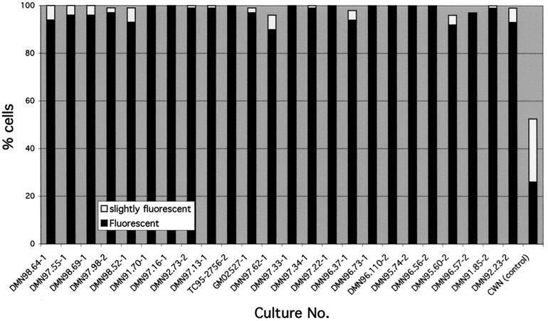
Mucolipidosis IV (MLIV) is an autosomal recessive disorder of unknown etiology characterized by severe visual impairment and psychomotor retardation. Recently, there has been considerable interest in positional cloning of the MLIV gene. It is unknown whether MLIV is a genetically homogenous disorder. In this paper, we present experiments that determined whether the MLIV phenotype in fibroblasts could be corrected by fusing normal cells to MLIV cells and fusing fibroblasts from pairs of patients. All of our MLIV patients fulfilled several diagnostic criteria that we developed. In addition, we found high sensitivity to chloroquine in cultured fibroblasts from MLIV patients. We found that normal cells corrected the MLIV phenotype. Fusion products of normal and MLIV fibroblasts, but not MLIV fibroblasts themselves, were relatively protected against chloroquine selection. In addition, 74% of the normal-to-patient fusion products had reduced levels or total loss of MLIV characteristic autofluorescence. However, there was no complementation of the phenotype in fibroblast cultures from any of our MLIV patients, including those of non-Jewish ancestry. In fusion products of MLIV cultures from 24 patients, 90-100% of the cells remained autofluorescent. These results indicate that all of our known MLIV patients, regardless of ancestry or severity of the developmental defect, have a single mutated gene.
Figures


Similar articles
-
Identification of the gene causing mucolipidosis type IV.Nat Genet. 2000 Sep;26(1):118-23. doi: 10.1038/79095. Nat Genet. 2000. PMID: 10973263
-
Mucolipidosis type IV.Mol Genet Metab. 2001 Jul;73(3):197-203. doi: 10.1006/mgme.2001.3195. Mol Genet Metab. 2001. PMID: 11461186 Review.
-
The molecular basis of mucolipidosis type IV.Curr Mol Med. 2002 Aug;2(5):445-50. doi: 10.2174/1566524023362276. Curr Mol Med. 2002. PMID: 12125810 Review.
-
Cloning of the gene encoding a novel integral membrane protein, mucolipidin-and identification of the two major founder mutations causing mucolipidosis type IV.Am J Hum Genet. 2000 Nov;67(5):1110-20. doi: 10.1016/S0002-9297(07)62941-3. Epub 2000 Sep 29. Am J Hum Genet. 2000. PMID: 11013137 Free PMC article.
-
Mucolipidosis type IV: an update.Mol Genet Metab. 2011 Nov;104(3):206-13. doi: 10.1016/j.ymgme.2011.06.006. Epub 2011 Jun 16. Mol Genet Metab. 2011. PMID: 21763169 Free PMC article. Review.
Cited by
-
Lysosomal TRPML1 Channel: Implications in Cardiovascular and Kidney Diseases.Adv Exp Med Biol. 2021;1349:275-301. doi: 10.1007/978-981-16-4254-8_13. Adv Exp Med Biol. 2021. PMID: 35138619 Free PMC article.
-
Isolated ocular disease is associated with decreased mucolipin-1 channel conductance.Invest Ophthalmol Vis Sci. 2008 Jul;49(7):3134-42. doi: 10.1167/iovs.07-1649. Epub 2008 Mar 7. Invest Ophthalmol Vis Sci. 2008. PMID: 18326692 Free PMC article.
-
The role of TRPMLs in endolysosomal trafficking and function.Cell Calcium. 2015 Jul;58(1):48-56. doi: 10.1016/j.ceca.2014.10.008. Epub 2014 Oct 28. Cell Calcium. 2015. PMID: 25465891 Free PMC article. Review.
-
Autophagy in lysosomal storage disorders.Autophagy. 2012 May 1;8(5):719-30. doi: 10.4161/auto.19469. Epub 2012 May 1. Autophagy. 2012. PMID: 22647656 Free PMC article. Review.
-
Autophagic dysfunction in mucolipidosis type IV patients.Hum Mol Genet. 2008 Sep 1;17(17):2723-37. doi: 10.1093/hmg/ddn174. Epub 2008 Jun 11. Hum Mol Genet. 2008. PMID: 18550655 Free PMC article.
References
-
- Berman E R, Livni N, Shapira E, Merin S, Levij I S. J Pediatr (Berlin) 1974;84:519–526. - PubMed
-
- Amir N, Zlotogora J, Bach G. Pediatrics. 1987;79:953–959. - PubMed
-
- O’Brien J S. In: The Metabolic Basis of Inherited Disease. 6th Ed. Scriver C R, Beaudet A L, Sly W S, Valle D, editors. New York: McGraw–Hill; 1989. pp. 1797–1806.
-
- Livni N, Legum C. Exp Cell Biol. 1976;44:1–11. - PubMed
-
- Kohn G, Livni N, Ornoy A, Sekeles E, Beyth Y, Legum C, Bach G, Cohen M M. J Pediatr (Berlin) 1977;90:62–66. - PubMed
Publication types
MeSH terms
Substances
LinkOut - more resources
Full Text Sources
Other Literature Sources
Research Materials
